Isao Nagaoki*1, Keiji Tamura*1, Hiromi Mise*2, Takashi Fujii*1, Akiko Wakui*3, Marina Wayama*3
Transmission electron microscopy (TEM) is an essential tool for research and development in diverse fields such as medical science, biology, polymers, chemicals, and nanomaterials. To meet the demands of these different fields, the HT7800 series of 120 kV transmission electron microscopes has been developed.
This series consists of two different instruments. The HT7800 is capable of wide-field, high-contrast observations due to the design of its lens system, whereas the class-leading HT7830 allows high-resolution imaging.
The features of the HT7800 series together with application examples are introduced in this report.
Figure 1 shows an external view of the HT7800. Both models in the HT7800 series allow images to be viewed under room-light conditions on a fluorescent screen using a screen camera, as was the case with the previous HT7700 model.
In addition, the acceleration voltage can be varied from 20 kV to 120 kV. In the HT7800 model, the magnification can be varied from ×50 to ×1,000 in Low Mag mode and ×20 to ×600,000 in Zoom mode. The HT7830 supports a maximum magnification of ×1,000,000 in Zoom mode.
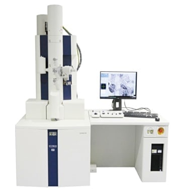
Fig. 1 HT7800 series TEM
The main features of the HT7800 series are described below.
(1) Ease of operation
The HT7800 series combines high performance with intuitive, user-friendly operation using a newly designed graphical user interface (GUI) and operation panel. Rapid TEM analysis with a minimum number of steps can be performed under room-light conditions using a high-speed screen camera. A newly developed pneumatic objective aperture (optional) eliminates the need to select the mode when changing magnification.
(2) Excellent image quality
In addition to the original Hitachi dual-mode objective lens, the electron optical system has been improved. In the HT7800 series, there are two models that can be chosen depending on the purpose of the observations; one is the HT7800, which allows high-contrast observations with a wide field of view. The other is the HT7830, which offers high-resolution observations.
In addition, the low acceleration voltage of 20 kV allows high-contrast observations of low-contrast specimens such as unstained tissue or carbon-based materials.
With the HT7830, an on-axis lattice resolution of 0.19 nm is guaranteed. Figure 2 shows a high-resolution TEM image of single-crystal Silicon (lattice spacing of 0.19 nm) obtained using the HT7830.
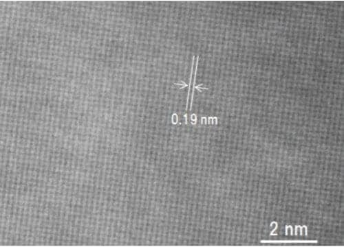
Fig. 2 Example of a high-resolution observation of a Silicon single crystal
Instrument: HT7830 Sample: Silicon single crystal Accelerating voltage: 120 kV
Magnification: ×1,000,000
(3) Improved functions
The HT7800 series offers a variety of automated functions as standard.
The HT7800 includes automatic 3D image acquisition with a specimen tilting angle of 70° to -70°, together with reconstruction algorithms.
In addition, both models have an Image Navigation function for locating a desired field of view. The Image Navigation function enables to search a desired area intuitively and obtain the image automatically by designating the area for acquisition.
(4) Versatility
A range of extensions are available depending on the user's needs, including cameras, and tomography, CLEM, STEM, and EDX systems.
Figure 3a shows an image of an unstained section of a rat ischial nerve acquired using the HT7800 at an acceleration voltage of 80 kV, in HC mode with a direct magnification of ×12,000. The image for myelin of rat ischial nerve section without applying electron staining can be observed with optimized contrast. An enlarged image of the region outlined in red is shown in Figure 3b. The layered structure of myelin can be clearly observed. Thus, the HT7800 can obtain high-contrast images of unstained sections.
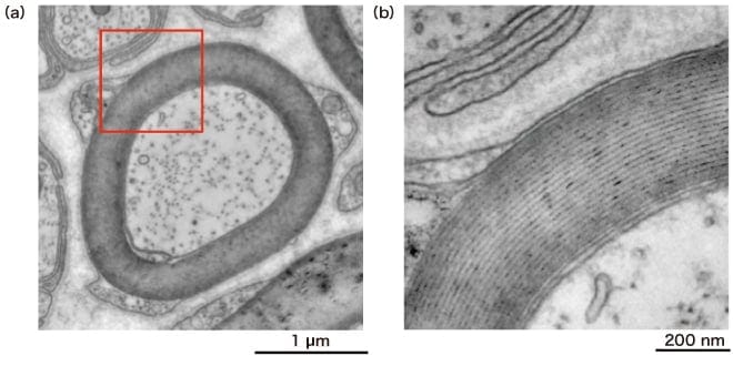
Fig. 3 Example of a high-contrast observation of an unstained fragment of a living organism
Instrument: HT7800 Sample: Rat sciatic nerve Accelerating voltage: 80 kV
Magnification: (a) ×12,000, (b) ×40,000
Figure 4a shows a high-resolution image of a fuel-cell electrode catalyst taken using the HT7830 at an acceleration voltage of 120 kV, in HR mode with a direct magnification of ×100,000. The distribution of platinum (Pt) particles on the carbon support can be observed. The region outlined by the red square is enlarged in Figure 4b. The lattice structure of the carbon support, with a spacing of 0.34 nm, can clearly be observed. Thus, the HT7830 allows high-resolution, high-contrast imaging of nano-composites, such as supported metal catalyst particles and carbon.
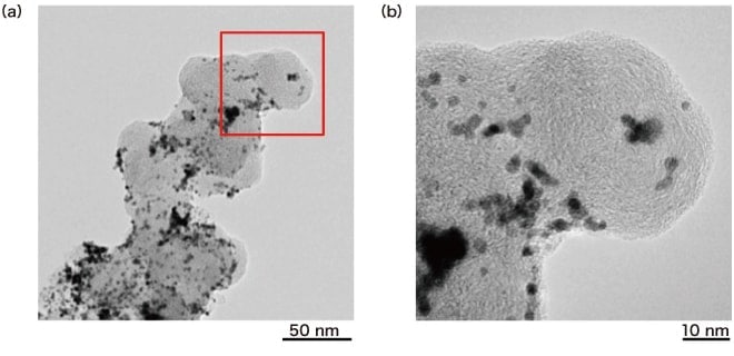
Fig. 4 Example of a high-resolution observation of a fuel-cell electrode catalyst
Instrument: HT7830 Sample: Pt/C catalyst Accelerating voltage: 120 kV
Magnification: (a) ×100,000, (b) ×400,000
The HT7800 series has been developed to meet the requirements of fields such as bio-medical science and nanomaterials. Like the previous model, the HT7800 series is equipped with a screen camera and a Hitachi original dual-mode objective lens. Moreover, in order to meet a wide range of needs, improved functions and enhanced operability are provided. The HT7800 for life sciences and the HT7830 for materials research offer high-contrast imaging and class-leading high-resolution performance, respectively. The HT7800 series microscopes are expected to be used as vital tools in various fields of screening, research and development.
About the authors
*1 Isao Nagaoki, Keiji Tamura, Takashi Fujii
Electron Microscope Systems Design 2nd Department
Science Systems Product Division
Science & Medical Systems Business Group
Hitachi High-Tech Corporation
*2 Hiromi Mise
Electron Microscope Solution Systems Design Department
Science Systems Product Division
Science & Medical Systems Business Group
Hitachi High-Tech Corporation
*3 Akiko Wakui, Marina Wayama
Application Development Department
Science Systems Product Division
Science & Medical Systems Business Group
Hitachi High-Tech Corporation
See more
