
Hideya Kawasaki
Doctor of Medicine
Chairperson
NanoSuit Research Laboratory, Division of Preeminent Bioimaging Research, Institute of Photonics Medicine,
Hamamatsu University School of Medicine
Histological investigations conducted with optical microscopy are essential in the life sciences and in medical diagnosis. Most formalin-fixed paraffin- embedded (FFPE) tissue sections are stained with hematoxylin and eosin (HE) for pathologists to view and make diagnoses. This process has been in place for over a century. Over the years, practitioners have accumulated extensive knowledge as they diagnose and classify diseases, and the number of banked tissue samples has grown to astronomical proportions1). The first immunohistochemical techniques were developed in the 1940s. Immunohistochemistry, which uses specific antibodies to find where different proteins are located, is now indispensable in the field of pathology. Optical microscopy allows rapid, convenient, and full-color observations, but suffers from relatively poor resolution.
The ultra-fine detail that transmission electron microscopes (TEMs) provide is critical for confirming diagnoses, staging disease, and predicting outcomes. Electron microscopy, however, has several disadvantages. Some of the reasons why electron microscopy-based diagnosis is not more widespread in clinical practice include the trimming, special staining, and other irreversible modifications of tissue sections required to acquire images with correlative light and electron microscopy (CLEM) from HE-stained samples, the need for specialist knowledge and techniques, the time required to imaging, difficulty identifying lesions, and difficulties in analyzing electron micrographs. There have been no techniques for non-destructive and reversible electron microscopy to allow viewing of the massive amount of FFPE, primarily HE-stained samples available throughout the world.
However, I discovered a way to use a biocompatible polymer solution (NanoSuit solution) and a scanning electron microscope (SEM) to non-destructively observe, at high resolution and in three dimensions, lesions in tissue sections identified through optical microscopy2). This technique opened the door to multimodal CLEM of identical sites in a given sample without damaging the sample. This paper discusses investigations conducted with the S-4800 field emission scanning electron microscope (FE-SEM) and the TM4000 low-vacuum scanning electron microscope (LV-SEM) by Hitachi High-Tech Corporation. It explores the possibilities of “seeking multimodal translational pathology from a single tissue slice.”
The large slide chambers of both SEM models that hold slides without breaking (cutting) them truly helped spur the development of the technologies I will tell you about.
With the NanoSuit technique, which the Hamamatsu University School of Medicine developed independently, a nanoscale membrane is formed for a short time around samples that can be non-destructively observed with a high-resolution SEM. Hariyama and colleagues discovered that exposing an adhesive extracellular substance that protects the body surface of fruit fly larvae to an electron beam or plasma enabled the SEM observation of live tissue under an extremely low pressure without sample drying3). Exposing extracellular substance to an electron beam or plasma polymerizes it, causing a nano-membrane to form that retains the gases and liquids in the organism inside in the presence of a vacuum.
Using a biomimetic approach, researchers selected biocompatible substances such as surfactants that mimic this extracellular substance and then polymerized the substances with plasma to form a nano-membrane on the surface of organisms. With this membrane, fine-grained surface structures and the movement of organisms can be observed using a SEM while preserving the organisms. Together with colleagues, I developed Surface Shield Enhance (SSE), whose active ingredient is glycerin4). SSE is biocompatible and works well with cells and tissues, preventing evaporation even when only a thin membrane is present. It is also electrically conductive. SSE therefore holds promise in different fields of research.
Lesions that pathologists identify in optical microscopy must inevitably be observed at higher resolution. High-resolution fluorescence microscopy has recently been used to analyze FFPE sections of colon cancer5) and breast cancer6) lesions. Fluorescence microscopy, however, requires fluorescent staining, making it incompatible with general HE-stained tissue. The electron microscope therefore remains an essential tool for high-resolution imaging. LV-SEM was recently used to analyze paraffin sections of primarily the kidneys. The Renal Biopsy LV-SEM Research Team is actively discussing how to proceed. In conventional techniques, SEM is not normally performed on existing HE-stained tissue slices. Rather, consecutive slices are irreversibly treated with gold, carbon, and osmium vapor deposition to impart electrical conductivity and then viewed with electron microscopy. Normal vapor deposition completely dries tissue slices, destroying their moist tissue structure. Moreover, when a vapor-deposited and dried tissue slice is again HE-stained, the coloration is altered, and these changes are practically irreversible. NanoSuit solution, however, offers ways to resolve these problems.
Pathologists working with a single tissue slice of a site of interest must spend substantial time and effort locating the lesion in the site because the SEM micrographs they use are high-resolution and monochrome. Assigning location information can help simplify the process. This is accomplished by locating the region of interest on a tissue slice, placing a mark on the back of the slide, and creating a digital micrograph of the lesion site with an optical microscope. The cover slip is removed with xylene for deparaffinization. Then, NanoSuit solution is applied, and the slide is spin coated to form a thin NanoSuit membrane. Once a membrane is formed, concentrated NanoSuit solution is applied dropwise around the site to mark it2). The mark allows the pathologist to easily locate the site of interest in high-resolution SEM. This process greatly simplifies CLEM observations of existing HE-stained samples (Figure 1A).
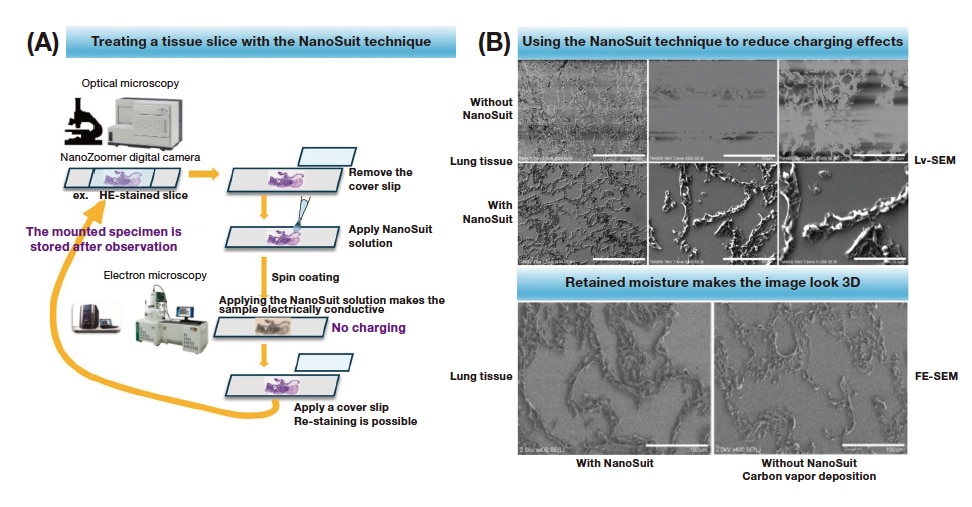
Fig. 1 NanoSuit-CLEM technique for observing FFPE slices
A: Procedures for NanoSuit-CLEM technique
B: Using the NanoSuit technique to reduce charging effects (LV-SEM) and micrographs of moisture-retaining tissue (FE-SEM) (secondary electron mode). Adapted from figure in Lab Invest. 2020;100(1): 161-173. Used under CC-BY-4.02).
A NanoSuit membrane confers electrical conductivity to tissue slices on microscope slides, which are insulators, thereby reducing charging effects. This allows clearer, more high-resolution SEM micrographs to be acquired. Notably, the tissue is imaged in its moist state (Figure 1B). Moreover, the marks made with NanoSuit solution can be non-destructively removed after SEM imaging to return the slice to its original state. Irreplaceable samples are thereby preserved. The optical micrographs produced in this procedure are two-dimensional color images that can be compared with three-dimensional electron micrographs taken of the same section. This technology was highlighted in a paper published after a related patent application was filed (Patent No. 7089756) and introduced as one of the “advances in translational pathology.”
Pathologists sometimes need a higher resolution view of sites they identify in optical microscopy. Viruses and even some bacteria are too small to be imaged as pathogens in optical microscopy. High-magnification observation with an electron microscope is required to view them. A technique with NanoSuit and CLEM was used to observe fungi, bacteria, and viruses.
First, specimens from patients with pulmonary aspergillosis were imaged with optical microscopy and FE-SEM. The images acquired were compared with those acquired with this NanoSuit-CLEM technique. The Aspergillus organisms and surrounding light eosinophilic fibrinoid substances seen under an optical microscope (Figure 2A) were clearly visualized in three dimensions with SEM in secondary electron (SE) mode. At greater magnification, the images showed sporangia, cell walls, septa, and nuclei in three dimensions (Figure 2B,C). Bacterial colonies can be difficult to identify (Figure 2D). Pairing NanoSuit with CLEM, however, produced images of bacterial clumps and groupings of cocci (Figure 2E,F). The microorganism was identified as Aeromonas hydrophilia in culture. The technique also provides a powerful way to identify virus particles too small to visualize with optical microscopy. In a patient with a cytomegalovirus (CMV) infection, many 100 to 300-nm particles (Figure 2H,I, white arrows), corresponding to CMV near giant-cell inclusion bodies forming in the kidneys, were found (Figure 2G).
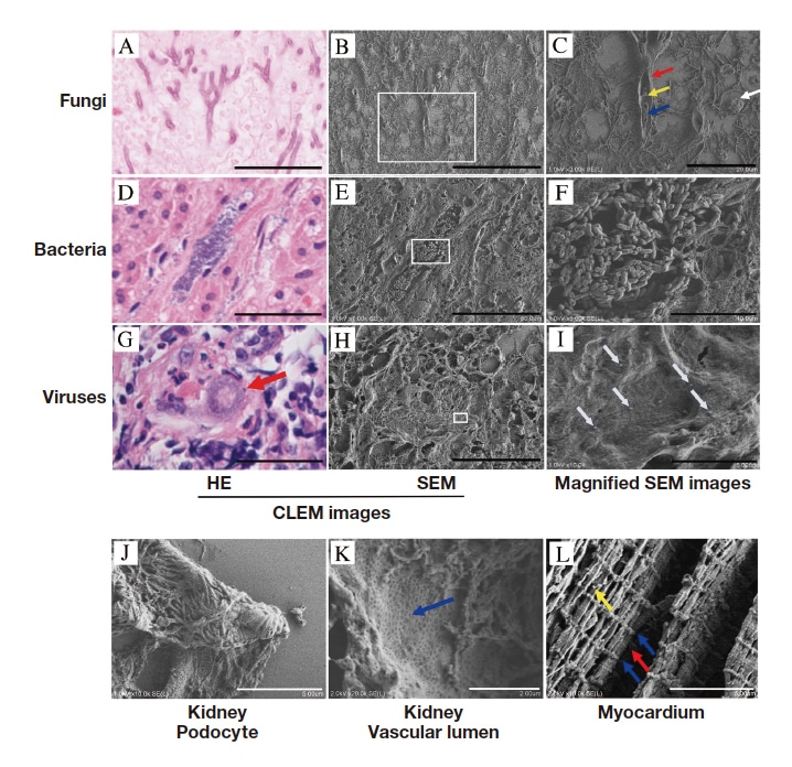
Fig. 2 CLEM high-resolution SEM micrographs of FFPE sections (secondary electron mode)
A-C: Pulmonary aspergillosis, A: HE micrograph, B: CLEM-SEM micrograph of A, C: Magnified SEM micrograph of the area in the square in B, white arrow: spore, red arrow: cell wall, blue arrow: septum, yellow arrow: nucleus. D-F: Bacterium (Aeromonas hydrophila), D: HE micrograph, E: CLEM-SEM micrograph of D, F: Magnified SEM micrograph of the area in the square in E. G-I: Cytomegalovirus (CMV), G: CMV inclusion body HE micrograph (red arrow), H: CLEM-SEM micrograph of G, I: Magnified SEM micrograph of the area in the square in H, with CMV particles (white arrows). J: Podocyte of renal glomerulus. K: Surface of renal vascular endothelium, blue arrow: indented area. L: Fine-grained structure in myocardial tissue with M line (red arrow), Z line (blue arrow), and mitochondria (yellow arrow). Adapted from figure in Lab Invest. 2020;100(1): 161-173. Used under CC-BY-4.02).
Pathologists also regularly need to observe fine-grained structures in lesions initially found with optical microscopy. In a previous paper, my colleagues and I discussed using the NanoSuit-CLEM technique to view fine-grained structures in a site of a tissue section initially observed under an optical microscope. Optical microscopy was unable to resolve these structures. We also clearly imaged podocytes in renal glomeruli (Figure 2J) and the uneven structure of fenestrated capillaries (Figure 2K). When we observed myocardial fibers, we saw the striated pattern in muscle fibrils (Z lines in I bands and M lines in A bands) and round mitochondria in three dimensions (Figure 2L). We also successfully clearly imaged in three dimensions protrusions from cells, vacuoles in cells, and fibers between tissues not viewable by optical microscopy. (These are not shown in the figures.) We also used immunohistochemical analysis and the NanoSuit-CLEM technique to show that some cells in human salivary gland adenoma7) and small cell lung carcinoma8) have primary cilia.
FFPE tissue sections are prepared through repeated cycles of fixation, paraffin embedding, deparaffinization, and dehydration. These steps can damage the fine-grained structures in some locations. But if areas of necrosis are avoided and other areas are sufficiently fixed and filled with paraffin, the fine-grained structures in those areas are preserved and viewable under a scanning electron microscope. The NanoSuit-CLEM technique could reveal changes in fine grained structures in a variety of diseases, leading researchers to new pathological mechanisms.
Immunostaining is a long-used tool for identifying areas of protein expression in pathology. The stain 3,3'-diaminobenzidine (DAB) is frequently used as a developer in immunostaining. Since metal chlorides and osmium tetroxide (OsO4) bind to DAB with high affinity, heavy metals can be used to enhance areas stained with DAB. NanoSuit membranes are relatively permeable to electron beams and therefore allow sites positive for DAB and metal chlorides or DAB and OsO4 to be visualized in SEM as high-contrast regions in backscattered electron (BSE) imaging.
My colleagues and I immunostained and DAB-developed structural proteins of human papilloma virus9,10), CMV9), varicella-zoster virus (VZV)9), and SARS-CoV-2 (unpublished) virus particles in FFPE slices. We then reacted 2% OsO4 with DAB and observed DAB/OsO4 positive regions in FE-SEM. Imaged VZV particles are shown in Figure 3. The skin blisters and subcutaneous areas of patients with a VZV infection were VZV-antigen positive (DAB positive) (Figure 3A). In SEM micrographs imaged with the NanoSuit-CLEM technique, DAB/OsO4 regions were enhanced as white regions in BSE mode (Figure 3B). Multiple white spots appeared in magnified regions (Figure 3C). Multiple particles were present in magnified images (arrowheads) (Figure 3D,E). No viral particle structures were visible in DAB/OsO4-negative regions. We concluded that these were VZV particles from their shape, diameter (150-200 nm), and electron-enhanced signals (arrowheads). Although osmium coating also visualized VZV particles (arrowheads), the contrast was somewhat low (Figure 3F). Multiple VZV particles are visible in CLEM-TEM micrographs corresponding to the micrograph in Figure 3C (Figure 3G,H). Secondary electron mode showed multiple independent VZV particles (arrowheads) on a blister on the skin’s surface (Figure 3I), whereas BSE mode (Figure 3J) showed DAB/OsO4-enhanced VZV particles (arrowheads) 9).
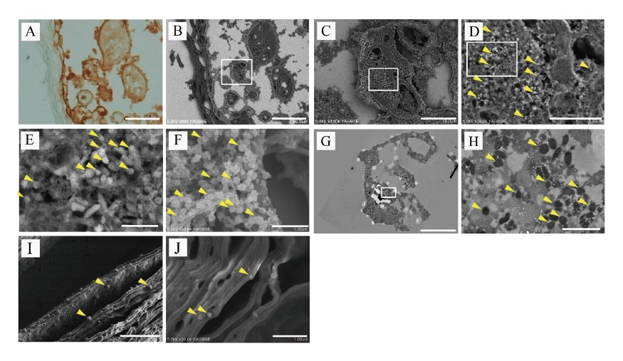
Fig. 3 CLEM micrographs of immunostained varicella zoster virus (VZV) samples
A: Optical micrograph of VZV-positive DAB-treated sample. B: CLEM-SEM micrograph of A (backscattered electron (BSE) mode). DAB regions (white) are enhanced. C: Magnified view of the square in B. D: Magnified view of the square in C. Virus particles (yellow arrowheads). E: Magnified view of the square in D. VZV particles (yellow arrows). F: Image of osmium coated sample. G: CLEM TEM image corresponding to the image in C. H: Magnified view of G. Multiple VZV particles are seen (yellow arrowheads). I: Multiple independent VZV particles on the epidermis (yellow arrowheads). VZV particles (yellow arrowheads) (secondary electron mode). J: Multiple independent VZV particles on the epidermis enhanced with DAB/OsO4 (yellow arrowheads) (BSE mode). Adapted from figure in Lab Invest. 2023;103(1): 100020. Used under CCBY-4.09).
A scanning electron microscope uses an electron beam to produce magnified images of sample surfaces. An energy dispersive X-ray analysis (EDX) instrument can be paired with a scanning electron microscope to perform elemental analysis of certain areas through the detection of characteristic X-rays. On identifying deposition of a foreign substance in HE-stained slices, pathologists sometimes predict what element or elements the foreign substance might contain and then apply a special stain. Such special staining, however, is labor-intense, requires experience, and is unsuited to identifying substances other than the one predicted. Scimeca and colleagues stated that EDX microanalysis could be a powerful tool for finding accumulated heavy metals in histological and forensic settings11). EDX performed with a conventional TEM and SEM, however, damages tissue slices, making re-evaluation infeasible. The NanoSuit-CLEM technique enables the concurrent analysis of multiple elements without destroying the sample2).
The following investigations serve as illustrative examples. EDX analysis was performed in a clinical setting on HE samples with suspected iron deposition. Areas with iron deposition were identified after HE staining (Figure 4A). Then, the NanoSuit technique was applied. Clear BSE images were acquired even with the electron beam at full (15 kV) in LV-SEM (Figure 4B). SEM/EDX analysis showed the brown area of deposition to contain iron (Figure 4C) and phosphorus (Figure 4D).
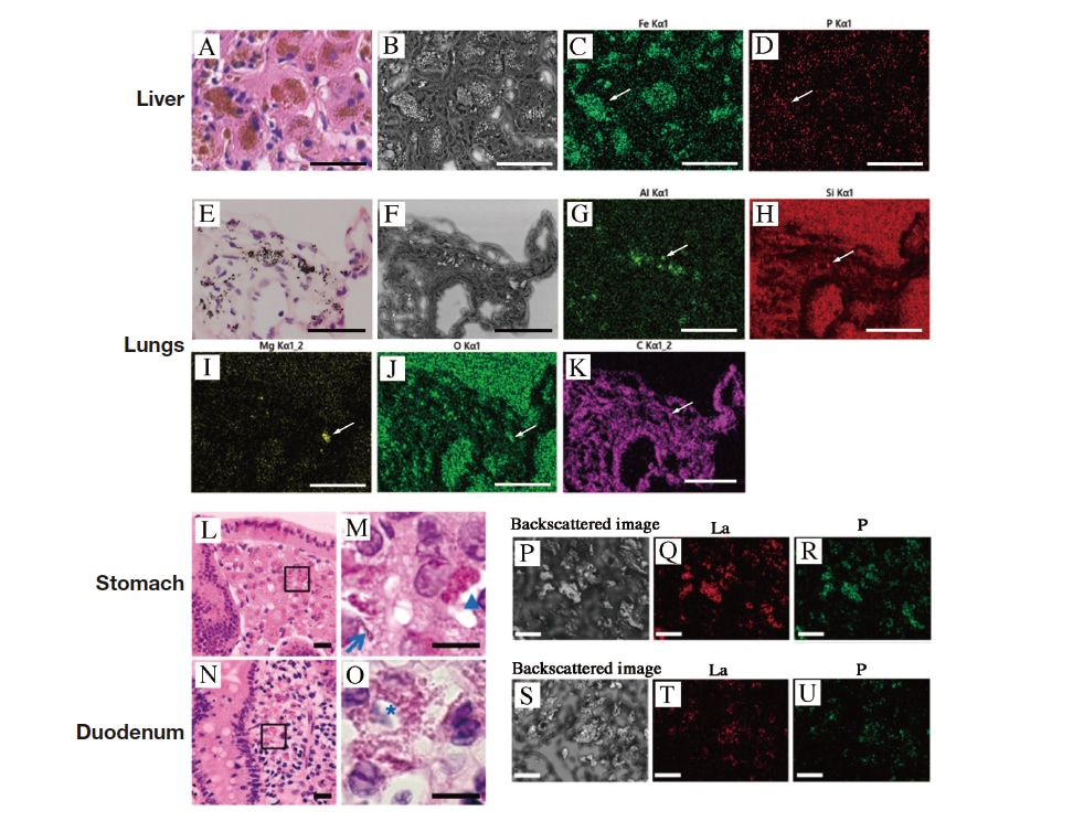
Fig. 4 Elemental analysis of FFPE slices
A: Area of deposition (brown), HE-stained. B: CLEM-SEM micrograph of A (backscattered electron (BSE) mode). C: Iron (white arrow). D: Phosphorus (P) (white arrow). E-K: Images of samples from a patient with black lung disease. E: HE-stained. F: CLEM-SEM micrograph of E (BSE mode), G: Aluminum (Al) (white arrow), H: Silicon (Si) (white arrow), I: Magnesium (Mg) (white arrow), J: Oxygen (O) (white arrow), K: Carbon (C) (white arrow). Adapted from figure in Lab Invest. 2020;100(1): 161-173. Used under CC-BY-4.02). L-U: Lanthanum phosphate deposition in the gastrointestinal tract. L and M: Micrographs of HE-stained stomach samples. N and O: Micrographs of HE-stained duodenum samples: Brown particles (asterisk), needle-like structure (arrow), and amorphous structure (triangle). P and S: SEM micrographs of areas with foreign matter deposition (BSE mode). Q and T: Lanthanum (La). R and U: Phosphorus (P). Adapted from figure in Diagnostics (Basel). 2019;10(1): 1. Used under CC-BY4.012).
In an analysis of a patient with black lung disease, multiple brown to black deposits were seen in HE-stained samples (Figure 4E). In BSE mode, strong backscattering signals arose from most deposits, but signals were absent in some (Figure 4F). SEM/EDX analysis of NanoSuit-treated samples showed multiple elements in lung tissue sections (aluminum (Al) (Figure 4G), silicon (Si) (Figure 4H), magnesium (Mg) (Figure 4I), oxygen (O) (Figure 4J), and carbon(C) (Figure 4K)), and many of the deposits of heavy metals present were oxidized.
Another investigation showed lanthanum phosphate deposition in the gastrointestinal tract12). HE-stained samples from a patient with suspected lanthanum phosphate deposition showed brown deposits in the shapes of particles, needles, and amorphous structures (Figure 4L-O). When the affected regions were treated with NanoSuit solution and observed with CLEM in BSE mode, regions with pigment deposition showed strong contrast (Figure 4P,S). Further study with elemental mapping using SEM-EDS showed lanthanum (La) (Figure 4Q,T) and phosphorus (P) (Figure 4R,U) localized in these regions.
A similar technique can be used to confirm deposition in the intestines of a calcium polystyrene sulfonate exchange resin used to reduce potassium levels in the blood (Kalimate powder, Kowa Pharmaceutical Company Ltd., Tokyo, Japan). This is done through the detection of sulfur in FFPE sections as one of the elements constituting Kalimate13). Given that deposition of Kalimate in the intestines can cause intestinal perforation and other serious complications, early identification and treatment discontinuation are critical. Research on patients who have undergone Helicobacter pylori eradication found black spots in the stomach, but patients with a current H. pylori infection rarely have black spots in the stomach. My colleagues and I used the NanoSuit technique with elemental analysis to observe these black spots, discovering that they consisted primarily of iron14). This discovery could further pathological knowledge of gastric disease and lead to the development of clinical tests.
As a pathologist, I frequently use these techniques in routine diagnostic work and find them helpful in characterizing foreign matter deposits in samples collected from organs. I am convinced they will become a powerful tool in pathology and diagnostics.
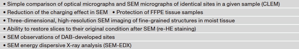
The NanoSuit solution developed independently at Hamamatsu University School of Medicine (1) enables clear SEM micrographs of fine-grained structures by forming a thin, hygroscopic membrane on slices to make them electrically conductive, (2) protects the tissues of organisms, such as mosquito larvae, enough to keep them alive when viewed under an electron microscope, and (3) is free of metallic elements that could interfere with the elements being viewed. The many unique features it provides are summarized in Table 1.
NanoSuit and associated techniques could help locate unexpected lesions with three-dimensional fine-grained structures in sites of CLEM-imaged tissue lesions found by pathologists or artificial intelligence and also provide alternatives to conventional diagnostic techniques performed with electron microscopy. The protection that NanoSuit membranes provide against damage from electron beams could be used in comparisons of single-sample CLEM with different analytical techniques. Patent application is pending as of March 2025. Advancing the possibilities of multimodal translational pathology from single tissue slices could uncover what is now hidden and help unlock the relationships between fine-grained morphological structures and molecular structures. NanoSuit stands to contribute to medicine as the new findings made with it help unlock disease mechanisms and establish new diagnostic procedures.
Table 1 Anticipated features of the NanoSuit-SEM technique used to observe FFPE slices
Acknowledgments
The author would like to thank the kind people at Hitachi High-Tech Corporation for their help with the research. The research was conducted with support from the Grants-in-Aid for Scientific Research (C) (20K07390).
References
See more
