Yusuke Ominami*1, Mari Sakaue*2, and Shinya Takaoka*3
Scanning electron microscopy (SEM), a technique used to characterize micromorphologies, has become a tool of essential importance across a variety of fields of research and development. Methods of atmospheric SEM (ASEM) have been reported to enable SEM observations of samples under ambient conditions, with a focus on water-containing samples such as soft materials or biological materials. The majority of existing ASEM techniques have been separatormembrane/ sample-contact ASEM methods, in which transmission of the electron beam is possible and the sample is placed in contact with a membrane to separate t he a mbient environment f rom vacuum. However, t his technique is problematic for observations of bulk samples, for which it is difficult to establish contact with the membrane. An additional difficulty of ASEM observations using this technique is that the membrane must be replaced each time the sample is observed.
For these reasons, we have devised an ASEM technique in which the sample is not in contact with the membrane: membrane/sample-non-contact ASEM. We have commercialized this technique for use with the AeroSurf 1500 atmospheric tabletop microscope, which can be used to carry out observations under ambient conditions (Fig. 1). In the AeroSurf 1500, the electron beam enters the membrane from the vacuum side and arrives at the ambient side through scattering and other processes. We use a membrane composed of silicon nitride (SiNx) to separate the ambient and vacuum regions. Our design allows the electron beam to arrive at the sample after scattering from ambient gas (Fig. 2). Our design also allows for the high-energy backscattered electrons to be retransmitted through the ambient region and membrane before arriving at the backscattered electron detector (Fig. 2). In this report, we discuss the features of the AeroSurf 1500 and present illustrative observations made using this instrument. We discuss our technique for correcting electron-beam scattering, which improves image distortion due to scattering of the electron beam between the membrane and the sample.
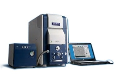
Fig.1 The AeroSurf 1500.
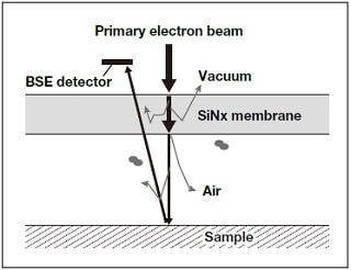
Fig.2 Principles of electron-beam detection.
Figure 3 shows diagrams of the internal design of the AeroSurf 1500.1) T he vacuum region of the AeroSurf 1500 includes a space within which ambient conditions can be maintained [Fig. 3(a)]. The interior upper wall of the ambient pressure chamber is equipped with a membrane that separates the ambient region from vacuum. This allows the sample chamber to be maintained at an ambient state (1 atm, or roughly 101,300 Pa), even while the remaining regions of the SEM interior are maintained at vacuum. To install a sample, one need only mount it on the sample stage and insert the stage into the chamber side. This establishes the sample in a position directly beneath the membrane [Fig. 3(b)]. Moreover, by using the vacuum pump provided as an accessory to the Aerosurf 1500, it is possible to maintain the sample in a low-pressure environment (from a few thousand Pa to 101, 300 Pa) [Fig. 3(c)]. Alternatively, by removing the membrane, it is possible to establish low vacuum conditions (a few Pa – few tens of Pa) [Fig. 3(d)].
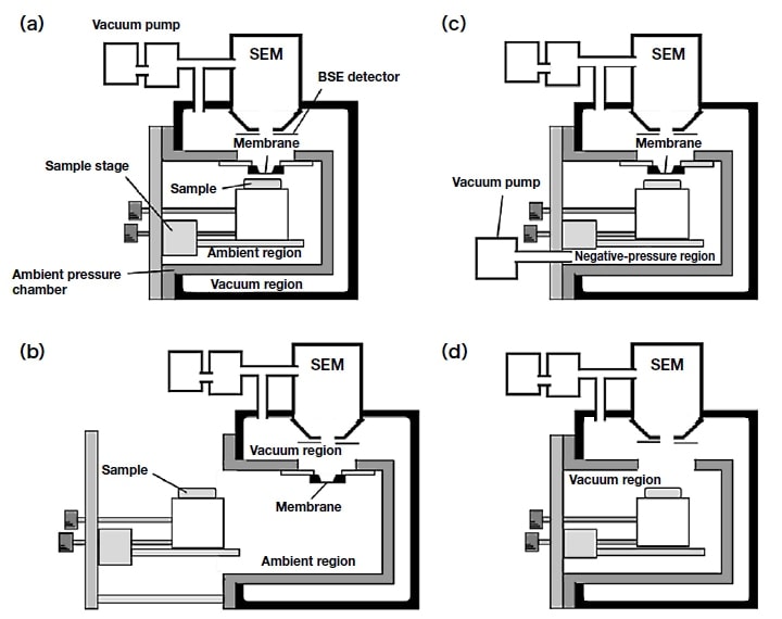
Fig.3 Diagrams of AeroSurf 1500 internal design.
Observations with the AeroSurf 1500 require that the sample be placed near the membrane. The relationship between the image quality and the membrane-sample distance is shown in Figure 4, where h denotes the membranesample distance and p denotes the pressure. At atmospheric pressure (101 kPa), scattering of the electron beam by the ambient gas results in poor image quality at separation distances of 100 µm or smaller; however, the image quality may be improved by reducing the pressure. The saturation vapor pressure of water is 2.3 kPa at room temperature, so watercontaining samples may be observed at pressures greater than this value without suffering water depletion. Therefore, successful observations may be achieved by reducing the pressure in the sample space as appropriate for the state and configuration of the sample.
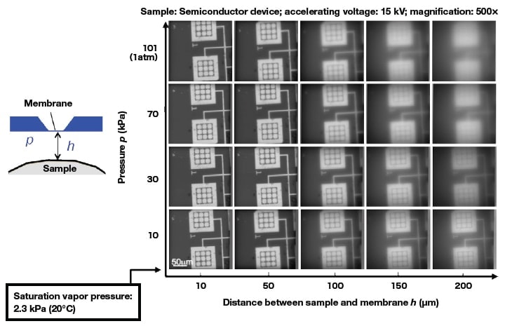
Fig.4 Relationship between image quality and membrane-sample distance.
Because the AeroSurf 1500 can be operated with the sample under ambient conditions, it allows SEM observations of morphology under conditions that are closer to the natural state and environment of the sample than is possible with conventional SEM techniques. Figure 5 compares images of a Japanese radish sprout captured under ambient conditions and in vacuum. Under ambient conditions, there is no evaporation of water due to vacuum suction, so the image here captures the sample in its natural water-containing state.
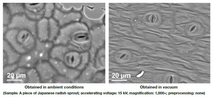
Fig.5 Results of AeroSurf 1500 observations.
Because the AeroSurf 1500 allows SEM observation of samples under low-vacuum conditions simply by removing the membrane, it allows visualization of the differences between the state of a sample as seen under ambient conditions and as seen in vacuum. Figure 6 shows images of the surface of a piece of pasta; here the same region of the surface is observed under ambient conditions and in vacuum. The image captured under vacuum conditions reveals cracks in the sample surface that are not visible in the image captured under ambient conditions. To check for artifacts in images obtained under ambient conditions, one may simply repeat the observation at higher resolution in vacuum.
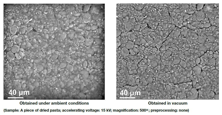
Fig.6 Results of AeroSurf 1500 observations.
Figure 7 shows the results of an experiment in which a cosmetic product was applied to a material, and the drying process was observed. At 30 seconds after application the sample retains a significant quantity of water. At 5 minutes the majority of the water has evaporated, and at 15 minutes the sample is nearly completely dried, as one can see by noting the large number of microparticles that have precipitated out of the sample.
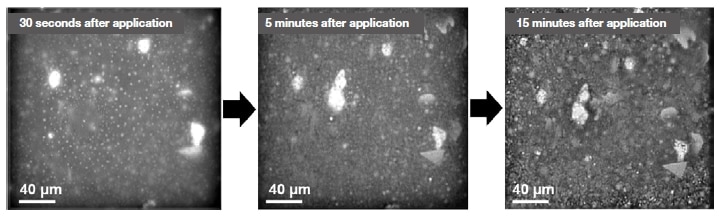
Fig.7 Results of observations of the drying process for a cosmetic product.
A standard accessory accompanying AeroSurf instruments is the ES-Corrector (electron scattering corrector)2) software developed for improving the quality of observations in cases where image quality has been degraded by electron-beam scattering in ambient conditions. By using this software, one can improve the quality of images captured under conditions ranging from negative pressure to ambient pressure. Figure 8 shows several images as captured under ambient conditions (above) and after improvement using the ES-Corrector (below).
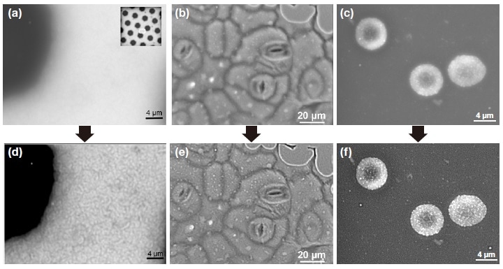
Fig.8 Images captured with and without ES-Corrector.
(a,d) Copper mesh. (b,e) Japanese radish sprout. (c,f) Rat red blood cell immunostained with gold colloids.
Accelerating voltage: 15 kV; room temperature; observed at 1 atm
With the AeroSurf 1500, it is now possible to make ASEM observations with the membrane and sample in a noncontact configuration. The ability to make observations under ambient conditions allows a variety of water-containing bulk samples to be observed in their natural state with no preprocessing. Moreover, ordinary low-vacuum observations can be made simply by removing the membrane. In addition, we have developed the ES-Corrector software for correcting the effects of electron-beam scattering in ASEM images. As we demonstrated, this allows major improvements in the quality of ASEM images. We are excited to offer the AeroSurf 1500 as a tool for imaging samples that could not previously be observed via SEM.
References
Author
See more
