Mai Yoshihara*1, Mitsuhiro Nakamura*2
In conventional scanning-electron microscopy (SEM), a sample for observation is placed inside a specimen chamber, which is evacuated to a state of high vacuum. The sample surface is scanned by an electron beam, and an image is acquired by detecting secondary electrons, backscattered electrons, or other signals induced by the irradiating beam. For samples that contain water, SEM observations typically require preprocessing steps such as drying or freezing the sample to prevent it from suffering shape distortions under high-vacuum conditions. However, there are a variety of situations in which it may not be desirable to observe a sample in a desiccated or frozen state, but rather to observe the dispersion and behavior of materials in liquids, as well as the morphology of living cells and microorganisms with the high resolution provided by SEM. This motivated the development of a liquid-sample observation technique using capsule-shaped sample holders to allow SEM observations while preserving sample solutions; these holders were formed from silicon-nitride membranes through which the electron beam could pass, and liquid samples injected into the interior of these holders could be maintained under atmospheric pressure. Observations using these holders employed high accelerating voltages and high-current electron beams capable of penetrating the membranes to irradiate the sample. The resulting transmitted and backscattered electrons were detected by detectors and used to generate images. However, this approach suffered from a number of drawbacks, including a high risk of sample damage due to electron-beam irradiation and difficulty in achieving high-contrast images for samples containing only light elements.
To remedy these shortcomings, Dr. Toshihiko Ogura of Japan's National Institute of Advanced Industrial Science and Technology (AIST) developed a new observation technique capable of imaging liquid samples without the need for high accelerating voltages or high electron-beam currents. Hitachi High-Tech then partnered with Dr. Ogura to develop a system for observing liquid samples, and this system—known as the Vitro detector—has been commercially available since 2023. In this paper, we first describe the principles of this new detector, and then present a series of sample measurements demonstrating its key advantages.
The Vitro detector comprises two components: the Vitro holder and a connector unit placed on the stage. Figure 1 shows a photograph of the Vitro holder and a schematic diagram of its cross-sectional structure. The liquid sample is sandwiched between two silicon-nitride membranes, the upper of which is coated by a layer of heavy metal; this upper membrane, together with O-ring seals, serves to isolate the sample from the vacuum environment, thus allowing the sample to be maintained under atmospheric pressure without drying. Positioned immediately beneath the lower membrane is a detector electrode connected to a preamp; this preamp, in turn, connects to the connector unit, from which the signal is acquired. To prepare a liquid sample for observation, the upper silicon-nitride membrane is turned upside down and droplets of the liquid sample are dripped on it from above. Then the lower membrane is put in place—sandwiching the liquid between the two membranes—and the assembly is turned right-side-up and mounted in the Vitro holder. This allows samples to be observed under high-vacuum conditions in their liquid state without drying.
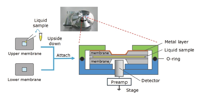
Fig. 1 Vitro holder (inset) and schematic diagram of its cross-sectional structure.
The signal-detection mechanism for the Vitro detector is illustrated schematically in Figure 2. Because the liquid sample is in contact with the lower surface of the heavy-metal-coated upper silicon-nitride membrane, applying a bias voltage to the heavy-metal layer creates an electric-field intensity distribution within the upper membrane that reflects the state of the sample. When the assembly is irradiated from above by a low-energy electron beam, the incoming electrons lose most of their energy due to scattering within the heavy-metal layer. Scattered electrons arriving at the silicon-nitride membrane then give rise to local variations in the electric potential, which are transmitted downward through the liquid sample and detected in the form of an electrical signal by the electrode beneath the lower membrane. The intensity of this signal depends on the electric-field intensity within the upper silicon-nitride membrane, and thus reflects the state of the liquid sample between the membranes.
Because this signal-detection mechanism does not require a high accelerating voltage or a high electron-beam current, samples observed with the Vitro detector are less likely to suffer damage due to electron-beam radiation. Moreover, because this approach does not require direct interaction with the sample, it is capable of producing high-contrast images, without staining or fixation, even for samples containing only light elements.
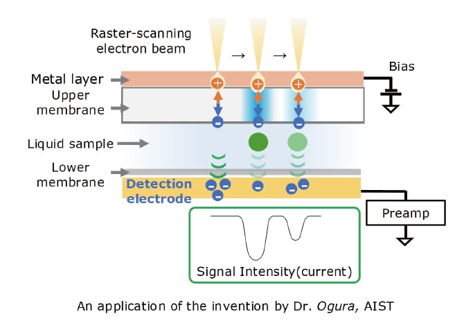
Fig. 2 Operating principle for Vitro detector.
We now present examples of actual measurements in which the use of the Vitro detector enables high-quality SEM imaging of liquid samples.
For milk and other dairy products, it has traditionally been difficult to obtain high-quality SEM images of samples in their natural liquid state. When attempting to capture images using backscattered electrons or other conventional signals, the damage caused by electron-beam irradiation has the effect of distorting the structure of milk fats as the scan proceeds. To illustrate how the Vitro detector solves this problem, Figure 3 shows SEM images of dairy products—specifically, milk (A,C) and powdered infant formula (B), observed using the Vitro detector (A,B) and the conventional secondary-electron detector (C) at magnifications of 10,000× (A-1,B-1,C-1) or 20,000× (A-2,B-2,C-2) 1). Samples for observation were prepared simply by injecting the dairy products in their natural liquid state into the Vitro holder. In images A-1, A-2, B-1 and B-2, white spherical structures with sizes of 150 nm to 3 μm are dispersed throughout the aqueous medium (w), and we interpret these to be milk fats (f). Images A-1 and A-2 also show an abundance of black particles with structures similar to those previously reported2); we interpret these to be casein micelles (c). A small number of black particles (p) are also visible in image B-2 for the powdered-milk sample. Because powdered milk is produced from milk containing casein protein, it is possible that these particles are also casein micelles. The smaller number of such particles in powdered milk may be evidence that the casein-protein content of powdered infant formula is intentionally reduced to levels similar to that of breast milk because it is difficult for infants to digest and absorb casein protein3,4). The clarity of the Vitro-detector images (A) is to be contrasted with the murkiness of the backscattered-electron images (C). Although the latter were captured from the same sample and at the same time as the Vitro-detector images, their quality is not sufficient to distinguish fatty components, casein micelles, or other structures.
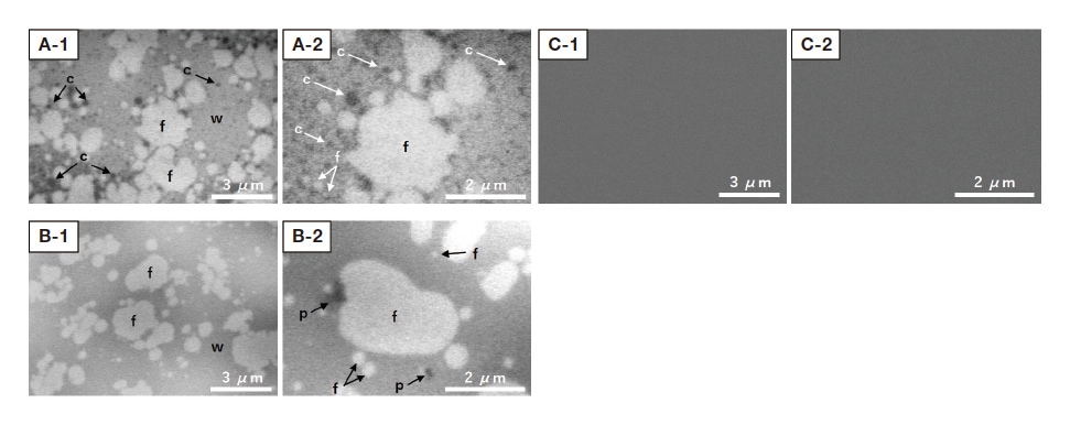
Fig. 3 SEM images of liquid dairy-product samples. A-1, A-2: Vitro-detector images of milk sample. B-1, B-2: Vitro-detector images of powdered infant-formula sample. C-1, C-2: Secondary-electron images of milk sample, captured simultaneously with the Vitro-detector images in A-1 and A-2. Instrument: SU5000. Accelerating voltage: 5 kV.
Magnification: 10,000× (A-1, B-1, C-1); 20,000× (A-2, B-2, C-2).
f: Milk fats. w: Water. c: Casein micelles. p: Particles.
Our next example involves SEM observation of liposomes. Liposomes are vesicles formed from phospholipids whose ability to encapsulate pharmaceutical substances or other active ingredients makes them promising candidates for applications to drug-delivery systems. To date, electron-microscopy observations of liposomes have employed techniques such as cryotransfer or negative staining5-7). Figure 4 shows an SEM image of liposomes captured using the Vitro detector. Note that this approach can successfully image liposomes of many sizes—from tens of nanometers to 500 nm—dispersed in a buffer liquid. Other types of biological samples have been studied by Dr. Ogura, who successfully observed liquids containing photosynthetic bacteria, protein antibodies, cultured cells, and other specimens8,9). As noted above, a key advantage of the Vitro detector is its ability to achieve high-contrast observations—even for biological samples containing only light elements—without staining or fixation.
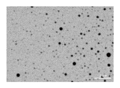
Fig. 4 SEM image of liposomes in liquid. Instrument: SU5000.
Accelerating voltage: 3.5 kV. Magnification: 10,000×
Our final case study is an observation of a cosmetic-lotion sample. Cosmetic lotions are typical examples of emulsions; like the dairy products discussed above, emulsions contain abundant oil components that make them highly susceptible to structural distortion under electron-beam irradiation. Figure 5 shows a Vitro-detector SEM image and energy-dispersive X-ray spectroscopy (EDS) mapping results for a sunscreen sample prepared simply by pouring the liquid sunscreen into the Vitro holder10). In the Vitro-detector image in Figure 5A, oil components (o), water (w), small particles (sp), and large particles (lp) can be observed. Figure 5B shows the corresponding EDS mapping results for the same field of view, obtained using an EDS detector (QUANTAX FlatQUAD, Bruker) positioned directly above the sample. The EDS analysis detects carbon (C) in oil components, oxygen (O) in water regions, zinc (Zn) in small particles, and titanium (Ti) in large particles. This example demonstrates that the combination of the Vitro detector and an EDS detector can yield high-quality SEM/EDS results for samples in their natural liquid state with minimal sample damage even for emulsions or other substances susceptible to damage under electron-beam irradiation.
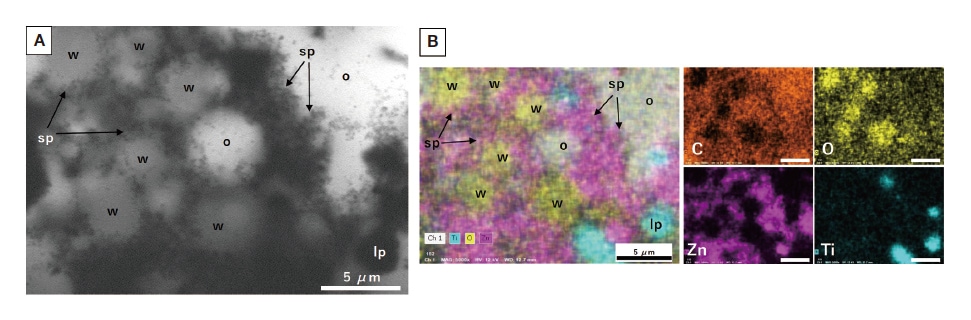
Fig. 5 SEM/EDS observation and analysis of sunscreen lotion. A) Vitro-detector image. B) EDS mapping image. SEM: SU5000. EDS: QUANTAX FlatQUAD (Bruker).
Accelerating voltage: 12 kV. Magnification: 5,000×.
o: Oil components. w: Water regions. sp: Small particles. lp: Large particles.
The Vitro detector is a new type of SEM detector now available for use with Hitachi SEM systems. This article described the operating principles for this detector and presented observation case studies demonstrating its unique capabilities. Key features of the Vitro detector include the following:
Going forward, we expect the Vitro detector to yield valuable new insights that will drive advances in the observation and analysis of liquid samples in the fields of biology, food science, and materials science.
Acknowledgments
In writing this manuscript, and in obtaining the experimental data presented above, we received invaluable suggestions and guidance from Dr. Toshihiko Ogura of the AIST Health and Medical Research Institute, to whom we extend our deepest gratitude.
References
About the authors
*1 Mai Yoshihara
Solution Development Department
Beam Technology & Analytical Systems Product Division
Core Technology & Solutions Business Group
Hitachi High-Tech Corporation
*2 Mitsuhiro Nakamura
Control Systems Design Department
Beam Technology & Analytical Systems Product Division
Core Technology & Solutions Business Group
Hitachi High-Tech Corporation
See more
