
Helena Téllez Lozano
International Institute for Carbon Neutral Energy Research
(wpi-I2CNER), Kyushu University
Collaborating authors:
John Druce
Tatsumi Ishihara
John A. Kilner
Although the effi ciency and durability of electrochemical energy conversion devices such as solid oxide fuel cells (SOFCs) and electrolyzers (SOECs) are drastically affected by their surface and interfacial chemistry and morphology, it is only recently that more attention is being paid to their characterisation. The increasing demand to perform this characterisation on smaller and smaller length scales is intertwined with instrumental and experimental developments for 2D- and 3D-structural and chemical analysis of complex heterogeneous materials, shallow surface layers, interfaces, and particles on the microscopic (> 100 nm) and nanoscale (< 100 nm) level 1, 2). In the present article, we will discuss how recent developments in two ion beam based surface analytical techniques - Time of Flight Secondary Ion Mass Spectrometry, ToF-SIMS, and Low Energy Ion Scattering (LEIS) spectroscopy - are contributing to advances in understanding of functional oxides for energy conversion.
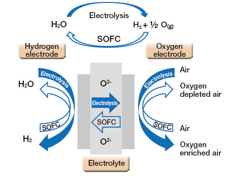
Figure 1.
Schematic of the operation of an oxygen-conducting SOFC-SOEC device.
Briefly, the electrochemical conversion of a fuel into energy (or the production of a fuel, such as H2, in electrolysis mode) in SOC devices (Figure 1) relies on exchange between the gas phase and the surface of the air electrode, as well as the subsequent ionic transport of the charge carrier (e.g. O2- or H+) through the solid oxide electrolyte towards the opposing electrode. These environmentally-friendly energy conversion systems typically require operation at intermediate (500-750°C) and high temperatures (750-1000°C), which can significantly reduce the lifetime of the cells. This degradation is due to additional processes such as poisoning (e.g. volatilization of metallic interconnects), segregation of impurities and other constituents, etc, occurring at the surfaces3). From this point of view, the insight provided by ion beam analysis techniques can lead to devices with improved behaviour.
In recent years, there have also been increasing efforts to determine the influence of the surface and near-surface chemistry on the catalytic activity of the electrode. Although the bulk diffusion properties in these functional oxides are, in general, well understood in terms of the bulk defect chemistry, there is as yet no atomistic description of the process of oxygen exchange between t he surface a nd t he gas phase. For instance, the surface exchange process is likely to be strongly dependent on the chemical composition and microstructure of the outermost surface, where the adsorption and incorporation of oxygen takes place (or oxygen evolution in the case of the SOEC). The results depict a complex mechanism in which impurities, secondary phases and other processes affecting the surface and near-surface defect chemistry might play a significant role that cannot be dismissed to understand the overall cell performance and durability4-8).
Analytical techniques utilising ion beams with energies less than 10 keV are well suited to the study of surface and near-surface composition because the penetration depth of incoming ions is much lower than that of electrons or X-rays used in other spectroscopic techniques such as Energy Dispersive Analysis of X-rays (EDAX) or X-Ray Fluorescence (XRF). The work we highlight in this article uses two different ion beam approaches, Time-of-Flight Secondary Ion Mass Spectrometry (ToF-SIMS) and Low Energy Ion Scattering (LEIS) spectroscopy, which provide information on the top ˜3 atomic layers and the very outer atomic surface, respectively.
In ToF SIMS, an energetic beam of ions (e.g. Bi+ at 30 keV in our ToF-SIMS V instrument (Ion-ToF GmbH., Germany)), referred to as the primary, or analysis, beam, is directed at the sample surface. The transfer of energy and momentum from the primary ion beam to the sample surface leads to the emission of secondary particles from the first 2-3 atomic layers of the surface (Figure 2(a)). The emitted particles consist of neutral and charged species, both monoatomic and molecular fragments, as well as electrons. Secondary ions of a chosen polarity are extracted into the analyser, and fly through a field-free drift tube at a fixed energy. This causes a dispersion of the ions on the basis of their mass-to-charge (m/z) ratio; lighter ions (e.g. H+, m/z = 1) have a higher velocity than heavier ions (e.g. La+, m/z = 139), and so arrive at the detector first. Hence by recording the number of counts versus the flight time to the detector, a mass spectrum of the extracted secondary ions is obtained. Because SIMS is a mass spectrometric technique, it can provide not only the elemental (and molecular) composition at the surface, but also the isotopic distribution.
In order to obtain high mass resolution, the primary ion beam must be pulsed into short bursts, resulting in low time-averaged currents of ˜ 1pA. However, this results in low rates of material removal . in fact, the analyses may be performed in a "static" regime, where the surface composition is not detectably changed during the measurement. In order to perform analyses of the compositional changes with depth, a dual beam approach is often adopted, in which a second low energy beam (e.g. Ar+ at 2 keV) is used with a higher current to sputter the surface.

Figure 2.
(a) Schematics of the LEIS and ToF-SIMS process.
(b) Detection limit and information depth of surface analysis techniques (Adapted from ref.[7]).
The second technique employed by our group to study the surface composition of solid oxide electrodes is Low Energy Ion Scattering (LEIS) spectroscopy. In contrast to SIMS, where we are analyzing the secondary particles generated by the impact of a primary ion beam, LEIS analyses the backscattering of the primary ion beam itself. In LEIS, a noble gas primary ion beam (most commonly 4 He+, 20 Ne+ or 40Ar+) with energy between 1 and 10 keV is directed onto the sample. Some of these ions are backscattered by binary inelastic collision with atoms in the sample, with a kinetic energy governed by the kinematics of the scattering event (scattering angle and mass of the surface atom). Thus the kinetic energy distribution of primary ions scattered through a known angle corresponds to a mass spectrum of the sample surface.
Because the projectiles are noble gas ions, the probability that they will become neutralized during the collision with the sample surface, and hence not detected by the electrostatic energy analyser, is rather high. Similarly, the neutralization probability of ions penetrating beneath the surface is virtually unity. Therefore, only ions scattered by the very outer surface atoms are detected, and the technique provides information on the elemental composition of specifically this first surface atomic monolayer9, 10). With these two techniques, we can obtain complementary information to provide powerful insight into the surface composition and reactivity.
The predominant application of SIMS in the field of solid state ionics in the last few decades has been for the determination of the mass transport kinetics of solid oxide electrode materials11-13). In particular, the use of stable isotope tracer (e.g. 18O, 2H) protocols, coupled with depth profiling analysis by SIMS is routinely applied to directly determine the kinetics of gas-solid exchange and bulk diffusion of the ionic carriers in SOC materials (e.g. oxygen or protons).
One of the advantages of isotopic tracer experiments when combined with SIMS depth profi ling is the capability to directly measure the ionic transport within the material. For solid oxide electrode materials which often show partial electronic conductivities orders of magnitude higher than the partial ionic conductivities (due to the higher mobility of electronic defects), isotope diffusion studies separate the electronic and ionic contributions far more readily than electrical measurements. Because it is a direct observation of the mobile ions, isotope labelling-SIMS studies also provide unequivocal differentiation between mobile species (e.g. O2- vs. Na+ in fast ion conductors14)). Furthermore, unlike like other methods such as relaxation techniques or electrochemical impedance spectroscopy, which measure the transport kinetics averaged at the macroscopic scale, this ion beam based methodology can extract microscopicallyresolved information of the active sites for oxygen incorporation giving its sub-micrometric lateral resolution, allowing the study of diffusion both in the bulk, and along and across grain boundaries and the exchange rate at the gaselectrode interface11).
This capability is illustrated in Figure 3, showing chemical and isotopic maps obtained for a composite ceramic material comprising Ce0.9Gd0.1O1.95 (CGO) and La0.6Sr0.4Co0.2Fe0.8O3-δ (LSCF) oxides15). The distribution of the phases is shown by the map for m/z = 88 u (fi g. 3c), corresponding to FeO2- and Sr-, both indicative of the LSCF phase . the bright regions in Figure 3c correspond to LSCF, whilst the dark correspond to CGO. The oxygen isotope maps for 16O- and 18O- secondary ions (Figures 3a and 3b respectively) can be used to obtain the 18O isotopic fraction ([18O-]/([16O-]+[18O-])) for each pixel (Figure 3d). This 18O isotopic fraction map shows similar microstructural contrast, with a heterogeneous distribution of the isotope fraction map for the LSCF and CGO regions.
Careful analysis of the isotope fraction map revealed that the regions appearing faster for oxygen exchange (with higher 18O isotope fractions in Figure 3d), in fact correspond to the CGO phase (no intensity in the m/z = 88 u image characteristic of the LSCF phase). This is contrary to what would be expected from the parent materials. LSCF is considered to be more active for oxygen exchange. This insight is possible only from the application of high lateral resolution ion beam analysis.
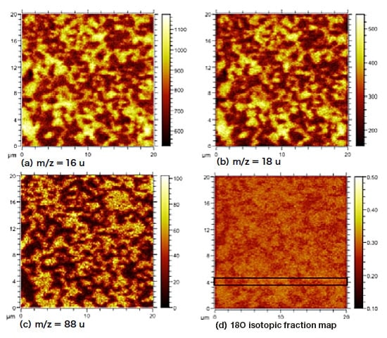
Figure 3.
ToF-SIMS negative secondary ion images of CGO-LSCF composite exchanged at 700°C.
(a) m/z 16 u (16O-) image,
(b) m/z 18 u (18O-) image,
(c) m/z 88 u (LSCF: FeO2- image,
(d) oxygen isotopic fraction map. Reproduced from Ref.15) with permission from the Royal Society of Chemistry.
On the other hand, measuring oxygen diffusion profiles in oxides by SIMS presents some challenges; the high negative secondary ion yields of oxygen at matrix levels can easily lead to saturation and nonlinearity of the secondary ion detection system, resulting in systematic errors in the measured isotope fraction. In conventional approaches, the use of Poisson dead-time correction and/or an overall decrease of the target current is typically employed to avoid nonlinear response of the detector16, 17).
Recent instrumental developments have improved the accuracy and precision of oxygen isotopic fraction measurements in solid material using ToF-SIMS by extending the linear dynamic range of the measurement by almost two orders of magnitude18, 19). This is a chieved by selectively attenuating the secondary ions with high useful ion yields and/or high concentrations that might lead to detector saturation (i.e. oxygen in the present example).
By using this technology, referred to as "Extended Dynamic Range" in the latest generation of ToF-SIMS V instruments18), the oxygen isotope fraction arising from short-circuit diffusion pathways (e.g. pipe dislocations or grain boundaries) in electrode materials can be reliably determined 19). As observed in Figure 4A, the use of high target currents and selective attenuation of the 18O- secondary ions at different levels required within the depth profile allows systematic errors derived from the non-linear response at the beginning of the profile where the 18O- intensity is above the conventional linear detection range to be avoided, while reducing the statistical uncertainty associated to the low intensities of 18O- at the end of the profile when the isotopic fraction approaches.
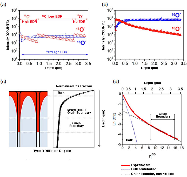
Figure 4.
(A) ToF-SIMS depth profile of the oxygen isotope distribution in an 18O-exchanged La0.8Sr0.2MnO3 ceramic sample by applying the selective attenuation of the secondary ions (SASI) approach to avoid detector saturation. The different attenuation factors applied during the depth profile are indicated by the arrows. (B) Corrected ToF-SIMS intensities of the oxygen isotope distribution profile according to the attenuation factors and Poisson correction. (C) Sketch of the Harrison type B diffusion regime allowing to distinguish the different bulk and grain boundary contribution to the 18O diffusion profile (Adapted from ref.12)). (D) Normalised 18O isotopic fraction (C'x) profiles as estimated from the ToF-SIMS profile. Two different contributions to the overall 18O diffusion profile are identified: diffusion through the bulk and fast diffusion pathways along the grain boundaries. Adapted with permission from 19).
In this way, we are able to extract the kinetic parameters governing the isotopic oxygen exchange (self-diffusion coefficient D* and surface exchange coefficient k*) with better accuracy and precision. In terms of the diffusion process, two regions with different kinetics are identified in the normalised 18O profile (Figure 4C and 4D): the slower diffusion through the bulk of the material (Db*) and the fast diffusion through the grain boundaries (Dgb*). Note that even in the "tail" which arises from the fast grain boundary diffusion, the scatter in the data is minimal.
The materials typically used as electrodes in SOCs are complex layered oxides with a perovskite-related structure. Under the conditions these materials experience during intermediate and high temperature operation (600-1000°C), these layered perovskite materials often show segregation processes, leading to deviations in the chemical composition at the near-surface and outer-surface compared to the bulk composition.
Recently, the chemical composition of the surface, and its effects on the surface exchange reaction, which ultimately limit the performance of the electrode, is being recognised as a major factor that should be investigated with further detail. In this sense, LEIS is becoming a very useful technique to investigate the segregation processes taking place in electrode materials, as it is able to provide information specifically on the composition of the first atomic layer (i.e. the gas phase-solid interface where the oxygen exchange process takes place).
The selection of the analysis conditions (e.g. primary ion species and energy) depends upon the desired information10). For instance, a He+ primary ion beam is required for the analysis of light elements such as oxygen or other light impurities like Na, as the heavier noble gas projectiles are not scattered by these light elements. In contrast, for the analysis of the cation surface coverage in these perovskite materials, the use of Ne+ or Ar+ primary beam is more suitable due to the better resolving power for these heavier species. This is illustrated in Figure 5A, showing the improvement in the resolution for the cation species present in a GdBaCo2O5+δ polycrystalline sample.
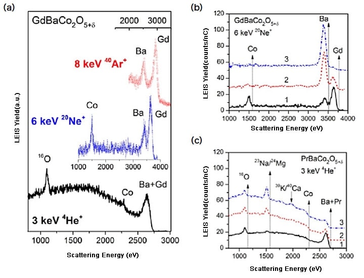
Figure 5.
(A) LEIS surface spectra representative of the bulk composition of a GdBaCo2O5+δ polycrystalline sample (as polished) as obtained with 3 keV He+, 6 keV Ne+ and 8 keV Ar+ primary ion beams. (B) 6 keV Ne+ LEIS surface spectra obtained from a GdBaCo2O5+δ polycrystalline as polished (1), after annealing in 0.2 atm O2 at 400°C for 1h (2) and after annealing at 1000°C for 12 h (3). (C) 3 keV He+ LEIS surface spectra obtained from a PrBaCo2O5+δ polycrystalline as polished (1), after annealing in 0.2 atm O2 at 400°C for 1h (2) and after annealing at 1000°C for 12 h (3). Adapted from Ref.5, 6, 20)
As mentioned previously, the high temperatures used during the fabrication and operation of a SOC leads to chemical and morphological changes at the surface of the complex metal oxides due to strong cation segregation and associated precipitation of secondary phases at the surface5, 6). Figure 5B shows a comparison of the chemical composition at the outer surface of the polycrystalline GdBaCo2O5+δ ceramic after different thermal treatments. Whilst the three cations are present at the outer surface after the polishing (Figure 5B.1), there is a rapid segregation of the Ba cations after annealing for 1 h at 400°C, with an associated decrease in the intensities for Co and Gd (Figure 5B.2). relatively short compared to the projected lifetimes of the devices. When the thermal treatment is performed at 1000°C for 12 h (e.g. conditions similar to those used for the sintering of the ceramic material), the surface is fully BaO-terminated with no other cation detected at the outer surface (Figure 5B.3).
These segregation processes have important implications for the oxygen exchange process between the gas phase and the electrode surface, as no transition metal cation, supposed to be electrocatalytically active species in these perovskite materials, is available at the gas solid-interface to participate in the oxygen reduction. Similarly, the presence of impurities in the ceramic material has a similar effect on the surface composition of the electrode at high temperature. For instance, Figure 5C shows a similar comparison of the surface composition for a PrBaCo2O5+δ sample. In this case, the impurities present in the ceramic material, coming from Pr precursors used for the synthesis, are susceptible to the same segregation process, hindering the detection of the Co transition metal at the outer surface. Therefore, the LEIS analysis of these surfaces provides information of the realistic surface where the oxygen exchange surface takes place under the SOC operation conditions.
The incorporation of oxygen from the gas phase into a ceramic electrode determines the performance of devices such as Solid Oxide Fuel Cells and electrolysers. The kinetics of this process is, in turn, dictated by the composition and morphology of the oxide surface. Recently, enabled by advances in ion beam analysis techniques, researchers are studying the chemistry of these functional surfaces.
Our group in particular is applying Time-of-Flight Secondary Ion Mass Spectrometry (ToF-SIMS) and Low Energy Ion Scattering (LEIS) spectroscopy. The combination of these techniques offers powerful opportunities to study the kinetics of mass transport and surface exchange processes (by ToF-SIMS combined with isotope labelling methodologies), in conjunction with the unique power of LEIS to study the composition of the very outer atomic surface of a sample – the same surface participating in the oxygen exchange reaction. The insight gleaned through these techniques promises to help unravel the complex chemistry and reactions at these oxide surfaces, facilitating the design of materials offering improved performance and stability for green energy conversion devices.
References
Collaborating authors
John Druce
International Institute for Carbon Neutral Energy Research (wpi-I2CNER), Kyushu University
Tatsumi Ishihara
International Institute for Carbon Neutral Energy Research (wpi-I2CNER), Kyushu University
John A. Kilner
International Institute for Carbon Neutral Energy Research (wpi-I2CNER), Kyushu University Department of Materials, Imperial College London
See more
