Powering Research with an Abundance of Ideas
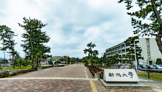
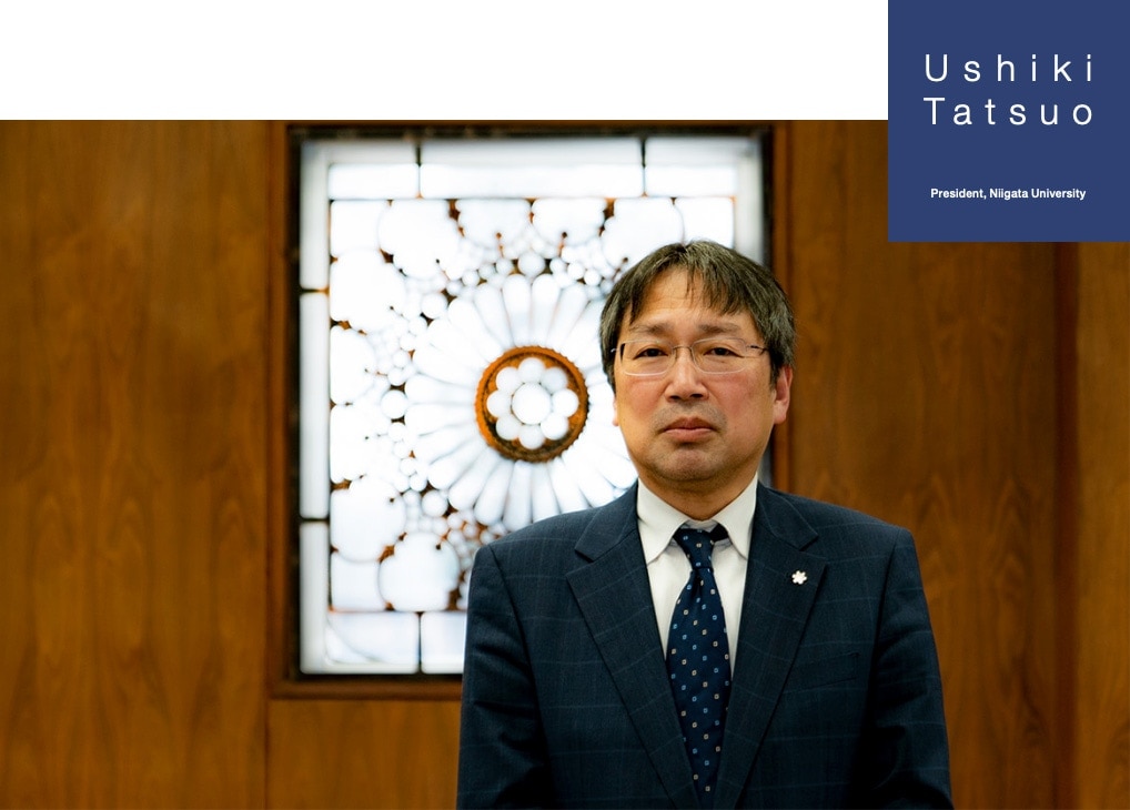
Professor Tatsuo Ushiki, himself a longstanding researcher in microscopic anatomy, is also the former chairman of the Japanese Society of Microscopy and is the current President of Niigata University since February 2020. I spoke with Professor Ushiki to look back on his research life and ask him about the progress he has made and his future expectations for his field.
Ushiki Tatsuo, President, Niigata University
The power of Tatsuo Ushiki’s paintbrush is known beyond just researchers, also reaching many other people through magazines and books for the general public. His realistic drawing of cells are full of mysteriously warm touches. Ushiki met with his mentor, Professor Tsuneo Fujita, through drawing and painting. It was Fujita who originally set Ushiki on the path to anatomical research.
Says Ushiki, “In high school, I was always in the art room drawing and painting pictures. When I decided to enter the Niigata University School of Medicine, my classmates were all amazed—they expected me to choose an art school. The reason why I went to medical school was because I liked living things. I wanted to enter a science or a medical school so that I could study biology.” At that time, the Japanese medical education program consisted of a two-year preparatory course (pre-med course), with students beginning studying specialized medical subjects such as anatomy and physiology from their third year. Professor Tsuneo Fujita was in charge of the lectures on histology. While the fact that Fujita’s lectures were a student favorite may also have factored into it, Ushiki became strongly attracted to the world of cells under the microscope.
Meanwhile, Professor Fujita had a strong passion for drawing and painting in addition to his professional academic pursuits. Every month he would invite models from an art model agency in Tokyo, to preside over a life-drawing club for students and citizens, as well as oil painting clubs. Before long, Ushiki was invited by his classmate to join the drawing club, which led him to visit Fujita’s laboratory. Here, he was also impressed by Shigeru Kobayashi, then an associate professor who would later go on to gain a professorship at Nagoya University. “With both of them, I was inspired by their passion for scholarship and painting, which naturally led me to decide studying in the lab as a PhD student.”
Professor Fujita was a great multi-talented scholar. In addition to drawing and painting, he also gathered students in the lab for a variety of reasons, like his early morning reading group on English, German, and French texts.
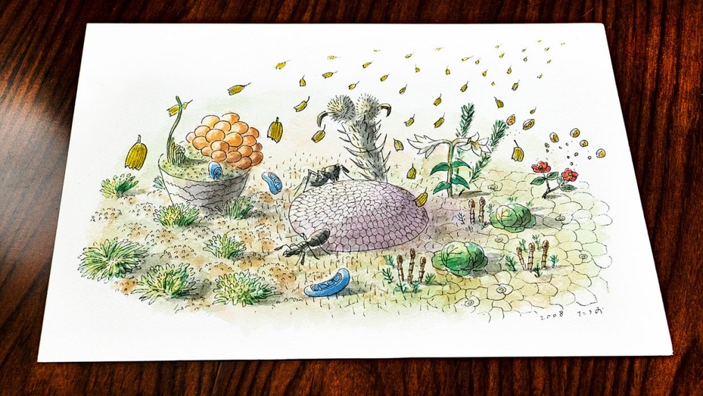
An illustration by Ushiki for a book in the Iwanami Junior Shinsho Paperback series, “Hidden Microscopic Wonders: Our Familiar World as Viewed Through an Electron Microscope” Ushiki depicts the structure as projected through an electron microscope like a flower garden
In 1978, about the time when Ushiki joined Fujita’s lab as a PhD student, scanning electron microscopes (SEMs) were starting to produce a variety of convincing results. Exactly
ten years prior in 1968, Professor Keiichi Tanaka of Tottori University became the first person in Japan to use an SEM to capture images of biological tissues. It was a micrograph of the lens fiber in the eye. “It may be the oldest SEM image taken in the world,” muses Ushiki. It was a landmark image, allowing direct 3D observation of the microscopic world in a state close to life, without the need to make a replica specimen as in the case with a transmission electron microscope (TEM).
Shortly thereafter, Keiichi Tanaka, Tsuneo Fujita, together with Junichi Tokunaga of Kyushu Dental University, organized an SEM symposium for medicine and biology, one of the first steps for SEM research in Japan.
In an SEM, as the generated electrons must avoid collisions with gas molecules before reaching the specimen, the interior is nearly a vacuum. Because of this, liquid cannot be introduced into the SEM, which usually means that specimens must be dry for observation. The lens fiber worked well because it was resistant to drying, but many specimens could not be dried without being deformed. At first, there
was a biting criticism that an SEM appeared to be viewing something that looks like dried fish.
One of the solutions to this was critical-point drying. Originally conceived of for TEMs, a paper that applied this method to SEMs became a hot topic at an SEM symposium. Based on these discussions, Tanaka commissioned a small factory to fabricate one. The process involves first putting dry ice in a chamber, letting it liquefy, and then applying heat and pressure until it becomes a gas. Fearing an explosion in the chamber, Tanaka reportedly watched from afar as the first experiment was conducted. Thanks to the dryer, however, the cells and tissues looked plump and full as if they were still alive. The experiments resulted in Hitachi’s HCP-1 criticalpoint dryer being built.
In 1972, Hitachi released a higher-resolution field-emission scanning electron microscope (FE-SEM), named HFS-2. It was at this point that biological SEM use really took off. Fujita’s most famous paper, published in 1974, was on SEM observations of the human spleen using the HFS-2.
From that point forward, SEM results accumulated. In 1980, Japan’s first international SEM symposium
(International Symposium on SEM in Cell Biology and Medicine) was held in Kyoto, with Tanaka and Fujita taking center stage. With the world’s leading SEM researchers in attendance, the symposium served as a valuable opportunity to show the world the high level of SEM research in Japan. Still a medical school student at this point, Ushiki was on hand as a helper and also got to attend delegation tours with the overseas attendees, in addition to other activities. “It was the first time I got to meet the esteemed Professor Radivoj V. Krstic of the University of Lausanne, known for his detailed 3D drawings of cells and tissues. Catching a quick glimpse of some of the world’s top researchers might have been another thing that started me down this path.”
By 1982, Ushiki had advanced to graduate school and started research in Fujita’s laboratory as a PhD student. This lab was established specifically for histological research and education on electron microscopy. In Fujita’s lab, studies performed with the already present TEM and SEM introduced by Fujita were mixed. Ushiki chuckles as he reminisces: “When I started to study in the lab, my main work was to replace the filaments, disassemble, and clean the TEM.”
As mentioned above, the first generation of SEM research was mostly accomplished at this point. Tanaka was further fighting to observe the internal structure of cells, for which specimen preparation proved elusive, whereas Fujita had pivoted his work from SEM research to histological studies on endocrine cells and paraneurons.
As Tanaka had found, the internal structure of a cell is difficult to observe. As nothing will be visible just by cracking the cells, observation requires removal of unnecessary substances to make only the desired structures stand out. In 1982, Tanaka’s lab stumbled upon a method for observing internal cell structures that involved dissolving some of the proteins in the cell with osmium tetroxide to expose just the membrane structure.
At the time, various digestion/maceration methods* started to be used for observation of the internal structure of both cells and tissues. Ushiki, who had been observing the structure of the thymus filled with thymocytes under an SEM in his PhD thesis, was subsequently transferred to Iwate Medical University as an assistant professor. There, he worked on various structures of the peripheral nervous system covered in a bush of collagen fibers. The goal was to obtain SEM images using a digestion/maceration method.
It was surprisingly difficult to remove the collagen components covering the nerve fibers. At that time, the hydrochloric acid (HCl)-collagenase method was used to selectively dissolve and remove collagen components. Trying various methods, Ushiki succeeded in dissolving only the collagen components without damaging other tissues using potassium hydroxide (KOH). With its high success rate, this superior method has become widely known as the KOH method.
Ushiki then moved to Hokkaido University as an associate professor, where he expanded upon such dissolution techniques and focused on SEM observations of various tissues, including osteocytes and osteoclasts. For example, to observe osteocytes covered in collagen fibers and calcium salts, just using the KOH method to dissolve collagen is not enough. Taking cues from observation methods for optical microscopy, he successfully observed cells clearly by first dissolving the calcium salts with a calcium chelating agent and then using the KOH method.
With his research analyzing the 3D ultrastructure of cells and tissues with an SEM using this series of techniques on track, Ushiki began working with an atomic force microscope (AFM) in an attempt to expand the possibilities.
The original AFM systems were based on the scanning tunneling microscope (STM), developed by Gerd Binnig and Heinrich Rohrer in 1982. In an STM, when the very sharp metal-tip (probe) is brought to a distance of about 0.1 nanometers from a metal specimen surface and a voltage is applied, a tunneling current flows through the gap. By monitoring the current and scanning the probe tip, a topographic image of the metal surface is created. Word about STM spread for its ability to take atomic level stereoscopic images at high resolution.
Then, in 1985, the AFM was developed. In place of a tunneling current, an AFM uses the van der Waals force between the tip and the specimen, allowing it to capture the topography of non-metallic surfaces. Later, this evolved into the scanning probe microscope (SPM). Ushiki was attracted to this technique, both because it allowed him to observe specimens without drying them and because it produced high-resolution images. Around 1990, he found a published paper on the successful observation of wet red blood cells using an AFM.
This could be it, he thought: maybe he could realize the dream of capturing 3D images of live specimens underwater at resolutions comparable to an SEM. Ushiki promptly made his appeal to the university to introduce an AFM system. His first attempt at imaging DNA well exceeded his expectations.
Triumphantly, he at last attempted to observe a cell. Doing so, however, proved not to be so easy. Live cells elude the probing tip when touched. Although he managed to succeed in obtaining images of chemically-fixed cells underwater, he was unable to capture images as he had imagined them. The cells became scratched or pushed by the probing tip, distorting their shape.
In 1995, Ushiki returned to the Niigata University School of Medicine as Professor of Microscopic Anatomy, replacing his retiring mentor, Professor Fujita. Unlike before, Ushiki was granted free access to the high-resolution FE-SEM and started his two-pronged research with both the SEM and SPM.
With his experience in graduate school disassembling and cleaning electron microscopes, he knew the structure and features of SEMs like the back of his hand. Along with his skills for making the most of the SEM’s capabilities, Ushiki also possessed a wealth of ideas on how to use it. In fact, as a graduate student, he was quick to focus on using backscattered electrons and was able to visualize lipid droplets of Ito cells in the liver by staining the cells with heavy metals and detecting them with backscattered electrons. He then wrote a paper on the findings he obtained.
Collaboration with manufacturers is essential for microscopic research, and Ushiki has been a prominent figure in the field since his joint research conducted with manufacturers in graduate school. Since becoming a professor, he has stepped up his collaborations even further with Hitachi High-Tech (formerly Hitachi Science Systems) and other manufacturers. This has allowed him to realize the ideas he had been percolating one after another, from improving the detector in low-vacuum SEMs to observing live plant cells without coating through a combination of an FE-SEM and a low-vacuum SEM. “I’ve had excellent levels of cooperation with everyone from engineers up to the president, with whom I’ve directly consulted on research. There have been times where we meet in the evening and our discussions started to take shape the next morning. Thanks to their support, SEM research has become one of the pillars of my work at Niigata University.”
Meanwhile, Ushiki’s AFM research, started back in his days at Hokkaido University, had evolved into leveraging the characteristics of AFMs to analyze the structure of collagen and chromosomes, partly in collaboration with Professor Mervyn Miles of the University of Bristol, who is a physicist distinguished for his groundbreaking new techniques with SPMs. One day back in 1998, Ushiki also noticed a paper on a scanning ion-conductance microscope (SICM) by Professor Yuri Korchev of Imperial College London. In it, Korchev used a glass pipette filled with an electrolyte solution instead of an AFM probe. Korchev had placed the specimen in a petri dish filled with an electrolyte, where one electrode was in the pipette and the other electrode was in the bath. He then applied a voltage between the electrodes, and measured the ion current flow through it to obtain the sample surface topography. The image shown was of the actual cell surface submerged in the liquid, and the image highly resembled an SEM image.
With his conviction on this technique, Ushiki went to meet Korchev, together with SPM expert Professor Futoshi Iwata of the Shizuoka University School of Engineering. In London, the two Japanese scientists were surprised to be shown a beautiful image of hair cells in the inner ear in liquid, similar to an SEM image. Upon returning to Japan, the two soon started building their own ion-conductance microscope. Although simple in principle and using what should have been the correct design drawings, they ran into problems with some of the finer technical details. During the time they fiddled with their own ion-conductance microscope, an overseas manufacturer released an AFM system capable of ion-conductance microscopy, making it available to them. Ushiki’s experience with the device led to improvements in the microscope under development. Ushiki would finally succeed in capturing cells and tissues in liquid. “At last, SEM-like 3D images can be taken in water. It only took 20 years!”
From the beginnings with optical microscopy, microscopes have developed and increased in resolution, through the electron microscope, probe microscope, and even superresolution microscopes. According to Ushiki, however, merely having a single high-resolution microscope is not enough; each microscope’s unique characteristics must be used in a complementary manner.
To do so, knowing your devices is important. “If you can get the most out of a successful combination of devices to find something interesting, then great.” While not always endowed with the best research environment, Ushiki got results by using the tools available to their fullest, and a bit of ingenuity. Ushiki believes there are still many possibilities for both SEM and SPM systems.
Where does Ushiki find the inspiration for his abundance of ideas? “It is important for people to come together and combine their wisdom. Walk through something together, and you’re bound to notice something new or something that you missed before. Even the most difficult problems can often be overcome by ideas.”
Over the 70 years since its inception, the Japanese Society of Microscopy, formerly chaired by Ushiki as its 58th chairman, has been a rare academic society in which researchers from both biological and engineering fields have participated. In today’s world of interdisciplinary research, cross-disciplinary collaborations and joint research are increasingly needed. The society also plays an important role in providing an opportunity for collaboration and for inspiring each other.
Ushiki’s outlook is also positive on his job as President at Niigata University. “It’s hard work, but I took office in an interesting time.” Founded in the Meiji Era as a national medical college, Niigata University has a long history and has grown into a large, comprehensive university, with two affiliated research institutes known as the “Brain Research Institute” that studies the human brain and its diseases, and the “Research Institute for Natural Hazards and Disaster Recovery” that investigates snowfall and other natural hazards. Ushiki feels it is necessary to envision a future where Niigata University takes advantage of such traditional strengths while also making the most of its regional characteristics. One example of this regionality is the recent creation of sakeology: the study of nihonshu, or sake. Such comprehensive academic fields fuse not only science, but also economics and culture. With the aim of taking Japanese sake culture to the next stage, they will also start offering a collaborative degree program with the University of Bordeaux in France.
Finally, I asked Ushiki if he had any requests for Hitachi High-Tech. “I want to see an SEM the size of a water bottle that is as easy to use as an optical microscope. It would be nice to be able to view its images on my smartphone. I’d love to be able to take out the SEM at the summit of Mt. Fuji if I get the sudden itch to experiment with something.” With that, he bears a boyish grin.
(Interview/article: Hiroko Hiratsuka)