A new approach to analyzing renal biopsy specimens
Using the distinctive characteristics of the low-vacuum scanning electron microscope (LVSEM), Dr. Sumire Inaga of the Department of Anatomy at the Faculty of Medicine of Tottori University developed a seminal new method for renal biopsy histopathologic analysis. The method represents a departure from the conventional perspective underlying the commonly applied transmission electron microscopy (TEM).
In this method, paraffin sections are prepared for light-microscope observation in three dimensions at the electronic microscopic level using the LVSEM.
Today, we invited its developer, Dr. Inaga, together with Dr. Nobuaki Yamanaka, an expert in renal pathology and nephropathy diagnosis and Director of the Tokyo Renal Research Center and Dr. Shinichi Okada, an Associate Professor in the Division of Pediatrics and Perinatology at the Faculty of Medicine of Tottori University, who has used the LVSEM to perform histopathologic analysis in collaboration with Dr. Inaga, to join us at the Tokyo Solutions Laboratory of Hitachi High-Tech Corporation to discuss the context and salience of the development by Dr. Inaga, as well as its future potential.
Sumire Inaga, Ph.D., Junior Associate Professor, Department of Anatomy, Faculty of Medicine, Tottori University
Nobuaki Yamanaka, M.D., Ph.D., Director, Tokyo Renal Research Center, Professor Emeritus, Nippon Medical School
Shinichi Okada, M.D., Ph.D., Associate Professor, Division of Pediatrics and Perinatology, Faculty of Medicine, Tottori University
The subjective symptoms of renal diseases do not emerge until the disease has reached an advanced stage, and they are generally regarded as difficult to classify. The structure and functions of the kidney are highly complex, and biopsies are particularly important for determining the relationship between disease progression and the morphology of the lesions involved. In the renal biopsy method developed in 1951, a small portion of renal tissue is excised using a slender needle and observed under a light microscope. This observation technique has since been complemented by renal immunohistopathology, in which information on immunologic reactions is obtained based on the presence and patterns of immunoglobulins, complement components, and other factors, and by electron-microscopic analysis. Today the information from all three sources is combined to distinguish diseases, determine the prognosis, and choose the appropriate therapy, and these special examinations are now performed routinely in renal biopsy analysis.
The use of electron microscopy in renal biopsy analysis has generated many new information. Most electron microscopic observations are performed using TEM, which requires a high level of knowledge and sophisticated techniques for sample preparation and image analysis as well as several weeks to obtain a definitive diagnosis.
Dr. Inaga's work in the Department of Anatomy at the Faculty of Medicine of Tottori University led her to wonder whether TEM, with these requirements, could really meet the needs of clinicians. This led to her desire for a simpler, faster method for conducting renal biopsy histopathologic analysis and ultimately to her development of the new method using the LVSEM. She had the opportunity to prepare biological samples for LVSEM for Dr. Kei-ichi Tanaka, who is a world-leading authority on SEM and the former professor of the lab. For, after his retirement from Tottori University, he changed the analytic tool from the ultra-high-resolution SEM with high-vacuum conditions to the low-vacuum SEM with backscatter electron (BSE) mode. LVSEM cannot reach TEM in terms of resolution, but enables the observation of samples that contain oils or water or are nonconductive. In addition, the LVSEM is relatively simple to operate and requires no special sample processing.
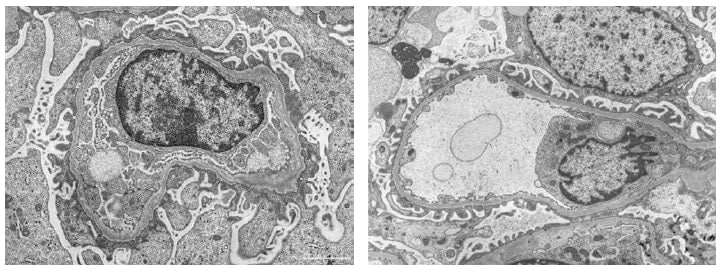
TEM images of renal biopsy specimens from cases of Alport syndrome (left) and thin basement membrane disease (right) are shown here for comparison of typical TEM images of these two pathologies.
Dr. Inaga took special note of these advantages. The starting point for her group's application to renal biopsy was the staining material. She notes her insight that "platinum-blue stain would become a keyword". It was a time of tightening limits on the use of uranyl acetate, the essential electronic stain. Platinum blue was gaining interest as a possible alternative, but none was available on the market for ready acquisition and use. Platinum blue was developed by Dr. Tanaka and his colleagues as a signal enhancer for LVSEM. Dr. Inaga proposed and patented the user-friendly stain kit that facilitated its utilization and led to its widespread adoption for TEM staining following reports at conferences and other venues of the highly effective differential staining of histopathologies by platinum blue in paraffin slices of rat- and mouse-tongue tissues, which are complex in composition.
When Dr. Inaga was further exploring the possibility of other applications, TEM personnel at Hitachi High-Tech Corporation suggested that renal biopsy sample would represent a particularly appealing application. The theory was that even though the observation would be by SEM, the kidney would be an appropriate target in view of the established method of diagnosis by TEM. Dr. Inaga notes that "As the doctor of pathology at Tottori University provided me with specimens that were from very representative cases, which enabled me to reveal the possibility of distinguishing of components in renal pathologic tissue using platinum blue."
On his first encounter with renal histopathologic analysis by LVSEM, Dr. Nobuaki Yamanaka, a leading expert in renal pathology and nephropathy diagnosis and Director of the Tokyo Renal Research Center, intuitively understood its importance as a new methodology and began a series of investigations into its potential uses.
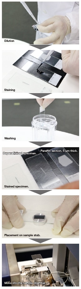
He soon discovered that applying the low-vacuum characteristics of LVSEM to the specimen by placing it on a slide glass in the sample chamber and observe it through the electron microscope made it possible to observe ordinary, light-microscope samples at the electron-microscope level, without the need for additional processing.
The combination of platinum blue with silver staining by PAM would heighten the analytical effectiveness of this method. PAM is well-established stain for analyzing renal biopsy specimens under a light microscope. As described by Dr. Inaga, "PAM stain is particularly advantageous for beautifully showing the component structure in the renal basement membrane. I noted that it was effectively the opposite of platinum-blue staining and realized these two stains could be used to obtain mutually complementary effects for observation."
She continues, "In particular, PAM staining has a long history, and Dr. Yajima, the former professor of Dr. Yamanaka's lab., improved PAM staining in Japan, which led to its present remarkable ease of use. PAM staining and platinum-blue staining are an excellent match, which is particularly advantageous for diagnosis of renal pathologies. I think the ability to see things with one that cannot be seen with the other and to use two specimens taken from the same tissue for comparative observation can bring greater depth to diagnosis."
Dr. Okada of the Faculty of Medicine at Tottori University, has worked in collaboration with Dr. Inaga in renal histopathology analysis, and in the perspective of the clinician on future possibilities for application of the LVSEM analysis method, he notes that "With an LVSEM, you can observe the renal biopsy tissue both at high magnification and in its entirety from corner to corner, as in a light microscope. That's an important advantage."
"In renal histological disease, lesions occur uniformly throughout the organ in most cases, but in focal segmental glomerulosclerosis (FSGS) and some other cases, the lesion initially occurs in one part before spreading to the entire organ. If you can't find that part and see the lesion, you can't recognize the abnormality."
"When using TEM, if an abnormality happens to be located outside the small part of TEM specimen under observation, then the diagnosis tends to be ‘free from abnormality’. But you don't really know whether abnormality is absent from the entire tissue. I think the capability of an LVSEM for observation of a wide area is very useful in examining for kidney pathologies in which the lesion begins in one part of the organ."
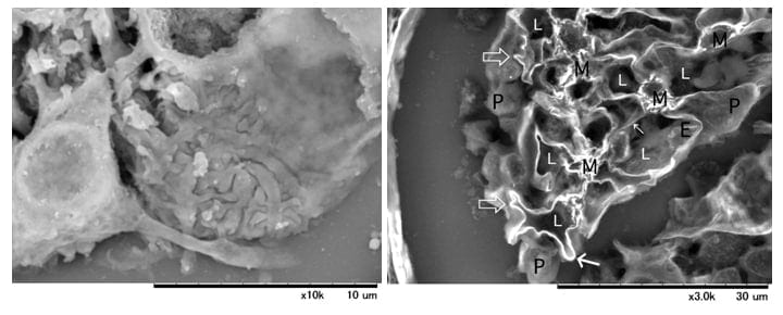
LVSEM images of a glomerulus in paraffin sections from a case diagnosed as FSGS, showing surface stained with platinum blue (left) and a cross-section stained with PAM (right).
Application of LVSEM is expected to both increase the amount of information that can be obtained from a single specimen and enhance its quality. It enables the 3D analysis of a wide range of morphologic abnormalities with both high resolution and high magnification.
Dr. Inaga emphasizes that "As might be expected, 3D images that can be seen stereoscopically under LVSEM, different from conventional TEMs and light microscopes, is very important." She notes "In cases of Alport syndrome, a mesh-work structure occurs in the basement membrane. Previously, diagnosis with a TEM was based on two-dimensional images of the section, and it appeared that the structural configuration might be basket-weave. With the 3D images, it became possible for the first time to really see that it actually is a basket."
Alport syndrome is a hereditary, progressive disease in which characteristic changes occur in the glomerular basement membrane. Similar abnormalities in the glomerular basement membrane can also be seen in thin basement membrane disease. However, in Alport syndrome, these abnormalities can progress to end-stage renal disease in youth, necessitating dialysis. It is therefore essential to distinguish between the two. As described by Dr. Okada, "The two are generally differentiated by conventional immunostaining and TEM and now also by genetic testing, but the results are sometimes inconclusive, and there is a strong possibility that LVSEM will be useful in this regard."
Dr. Yamanaka also points out the significance of obtaining 3D information, noting that "When you observe a sample through a light microscope, you are observing a sample that is 3 µm or in some cases 5 µm thick, but with an LVSEM, you can see a 3D image of the sample in its full thickness and ascertain aspects that you couldn't see before in 2D observation. Our real world is spatially 3D, and when you observe it in two dimensions, you are observing a world of lower dimension. When you see the 3D images for some cases, the differences from what you saw before in 2D are recognized clearly.
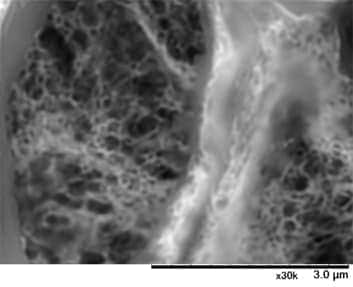
LVSEM image of a PAM-stained glomerulus from a case of Alport syndrome, clearly showing the mesh-work structure in the basement membrane.
Dr. Yamanaka has considered application of the LVSEM to tumor tissue diagnosis and cytologic examinations to detect cancer cells in pleural effusion and ascites fluids, with impressive results. To realize the full potential of LVSEM for renal biopsy diagnosis and nephropathologic research and its application to tissues other than renal and cases requiring fast, timely diagnosis, it will be necessary to ensure specimen quality and to develop and maintain the basic diagnostic criteria. It will also be necessary to ensure the capabilities of the image analyst to ensure the appropriate interpretation of the diagnostic pathology of specimens.
In the words of Dr. Okada, "With the LVSEM, calling up the image is easy. Seeing it there and thinking about it can be quite difficult. The first time I did it, Dr. Inaga was with me. She guided me one-on-one, pointing to objects on the screen and telling me which were cells, which were basement membranes, and so forth."
"Without that kind of guidance, it is probably hard to understand what you're looking at in the images when you see them for the first time. Things look somewhat different from what you're used to seeing with light microscopes and immunofluorescent methods. You can make out the images of things, and yet you can't tell what exactly they are. I think that in this sense, too, provision and publish of a database, such as 3D appearances of glomerular structure with epithelial cells, or that of IgA nephropathy for examples, and so forth, to develop the understanding, will be valuable."
Dr. Yamanaka has long advocated for the construction of an expanding database containing a large number and variety of cases in order to develop and disseminate an understanding of the LVSEM histopathologic analysis method and its important advantages.
In his words, "The basic categorization of renal diseases has to some extent been determined, but the relatively rare cases in which (these diseases and) their lesions have been observed is still small, and with small numbers, there is always a possibility that their occurrence is unusual to those cases."
"For the rare diseases such as Alport syndrome and thin basement membrane disease, the need is to gather many cases of each category and establish a database for making the correct diagnoses. This will require the broad distribution of LVSEM in use of the methodology."
Dr. Inaga observes that reading LVSEM images is not in itself a difficult problem and that the essentials can be readily mastered in seminars with demonstration units. With her own experience in its development extending over 10 years, she notes that the task of expanding recognition and utilization as a superior methodology remains in the future.
In closing, she notes that "Renal pathology is the most difficult of pathologies. Even if an outsider of pathology like me will easily perform kidney diagnosis using this method, there will be no actual progress.
"Thanks to Dr. Yamanaka's instruction and guidance in renal disease analysis and his introduction of this method in many venues, interest has grown among many. In a real sense, he has brought us to the starting line. In this spirit, it is my hope that an impressive volume and extensive range of data will be gathered, and that exact diagnostic criteria and methods will be identified and established, perhaps together with atlases or other forms of information, and widely disseminated."
This database is available in our membership website "S.I.navi" at the following URL.
See more
