Exploring Food Structure to Establish the Science of Cooking
Culinary science has a variety of approaches to research. Cooking methods, ingredient analysis, food properties, psychometrics (sensory evaluation), food culture, and food histology all comprise important parts of this research. Flavor and texture, such as hardness or chewiness, are all closely related to a food’s structure, and food histology has evolved along with techniques for observing tissue structure in order to examine the properties of each type of food.
For this interview, I sat down with Professor Machiko Mineki, who has been developing techniques for observing the microscopic structure of tissue, and has received many accolades. I asked Professor Mineki about progress in food histology research and what role electron microscopy plays in it.
Machiko Mineki, Tokyo Kasei University Professor, Department of Food and Nutrition, Faculty of Home Economics Doctor of Agriculture, Laboratory of Applied Gastronomy
Safe, balanced and delicious meals are the foundation for a healthy and comfortable life. The key to such meals is simple: cooking.
Starting with its definition, food histology involves visually determining the structure and materials present in fresh, cooked, or processed foods with the help of a microscope. Even the slightest change in the cooking process can produce a difference in taste, and food histology is considered a very powerful tool for determining what produces such differences.
Professor Mineki elucidates. “What methods will ensure that anyone can cook a delicious meal? And what makes it delicious? Answering these questions scientifically are the challenges of gastronomy. Food structure in particular provides us with important clues. Take pudding, for example. Pudding has such a smooth feeling in the mouth because of the amount of fat it contains and how well it is dispersed.”
Basically, this means that if we can clarify how the type of cream (animal-based or plant-based) and its arrangement impacts the tissue structure of the pudding, the taste of a delicious pudding can be reproduced.
“If we can determine the structure, it might wind up as data for food manufacturers to develop products, for example.”
What makes something taste good? How is good food made, and what can we do to make it taste better? As mentioned earlier, culinary science seeks to answer these questions. In Professor Mineki’s words, observing the tissue structure of food is visually appealing and is a method that makes the structure readily apparent to all.
“Structural analysis visualizes the structural features of a food that is associated with its physical properties, and makes changes in that food readily understandable for anyone. Culinary science is about researching taste, but researchers involved in structural observations always say how beautiful the structure of delicious food is.”
Delicious foods are structured beautifully. This goes to show that structure strongly correlates with taste. If the surface and internal structure of a food are different, the texture will differ. Composition also plays a part. The presence of underlying structure can also be determined by understanding differences in composition, such as through histochemical detection or X-ray analysis. Thus, by analyzing the structure of food, culinary science gives us more and more clues.
Professor Mineki’s 40-plus years of research has also focused on systematizing culinary science. For Professor Mineki, working as an assistant in the cooking laboratory of a women’s college was her first nudge down the path.
“When students in cooking classes would ask me why we were using a particular cooking method, I often couldn’t answer them immediately. It made me want some clarity on those questions.”
While not so common these days, at that time it was normal to add flour when boiling cauliflower, or to use saltwater when washing strawberries.
“But back then, no matter where I looked, I couldn’t find out why.”
At the time, chemical component analysis was often used as a way to answer these questions. Professor Mineki thought there had to be a more easily understandable way than component analysis—a way that anyone could understand.
“At the time, a professor from our neighboring food science laboratory taught me that if you dye bread dough, and then slice the bread baked from that dough, you can visualize the different ingredients. The images he then showed me using an optical microscope were beautiful and fascinating: the starch in the bread slice was dyed pink and the gluten was blue”.
That was the very moment Professor Mineki chose to study food structure. At first she studied bread, observing changes in lipids throughout the breadmaking process. After that, the university acquired a Scanning Electron Microscope (SEM), and Professor Mineki began observing the structure of various types of food. At this point, she was already comparing optical microscopy and SEM images. She learned how to prepare SEM specimens at a Japanese Society of Electron Microscopy (JSEM) seminar taught by Professor Masako Osumi of Japan Women’s University. She speaks of being blessed in terms of mentors.
“For many years, I was mentored by Pf.Emiko Matsumoto, a former professor at Kyoritsu Women’s University, who founded the Japan Society of Food Histology and introduced me to microscopy observations. At that time, Pf. Matsumoto would come back from trips with all the dishes she had eaten and was interested in, and she told us to observe them using SEM. Thanks to her, every week free time without class we would observe countless food samples, both animal-based and plant-based, over and over again. The observational skills Professor Mineki honed through repetition lead to further research.

Figure 1: SEM image of bread (Hitachi S4000) after fixation with glutaraldehyde and osmium tetroxide, drying, and ion-coating with Au. (Arrows indicate starch grains in flour gelatinized by heating)
Professor Mineki was advised to study for a degree on egg cooking at the Laboratory of Animal Morphology (now the Laboratory of Functional Morphology) at Graduate school of Agricultural Science, Tohoku University, which established the field of food histology.
“Studies on the structure of eggs began in the 1940s. Egg fixation*1 was quite difficult; especially for raw egg yolks, and research only got as far as viewing partially fixed egg samples.”
Professor Mineki had thought that both the overall structure and microstructure of eggs were well understood, but this was not the case.
“That was when we started viewing things under a Transmission Electron Microscope (TEM) in addition to an optical microscope and SEM. It was a first for me, and was really tough.”
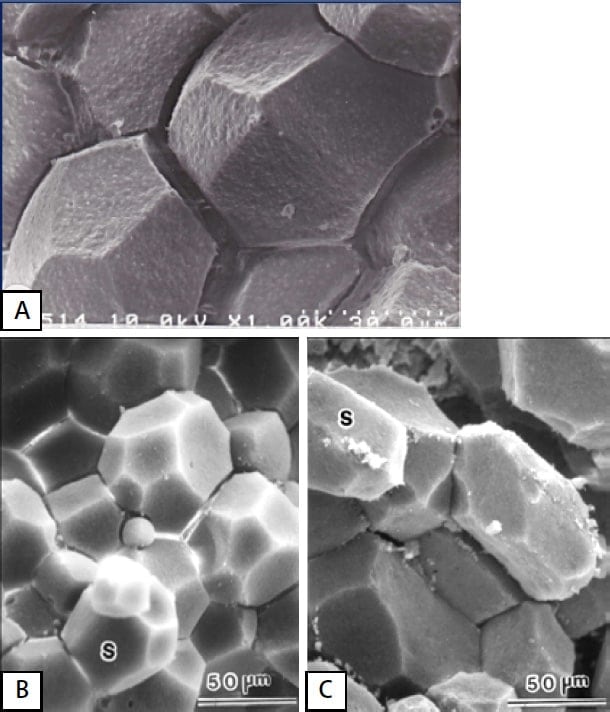
Figure 2: SEM images of egg yolk sphere
A: Yolk of fresh egg
B: Outer yolk of hard-boiled egg
C: inner yolk of hard-boiled egg
By first freezing the egg yolk with liquid nitrogen, dissecting it, and then performing fixation with a warmed fixing fluid, Professor Mineki was successful in observing the outer, inner and central parts (latebla) of a raw egg. The results were posted in the Journal of Food Science.
“When you observe an egg yolk sphere up close, it’s polyhedral, with different shapes in the center and ends of the egg. If the egg is physically impacted somehow, it affects the yolk sphere. Based on this, we determined the basic structure of the yolk in a raw egg. We also clarified the changes in tissue between fresh-laid eggs and eggs stored for a certain period, mainly using TEM. We clarified the differences in structure caused by heating while cooking, as well as the effects of seasoning.”
For this research, Professor Mineki received a Ph.D. in Agriculture from Tohoku University.
Professor Mineki speaks of the potential for hen eggs. “There is still plenty left to research about eggs. As a source of protein, eggs are relatively inexpensive and can be distributed at low cost. These qualities make them an important ingredient for countries and regions experiencing rapid population growth. Beef is said to have high loss in terms of feed efficiency. Behind insects, eggs are the second richest protein source. Hen eggs have widespread applications: they can be adapted to any kind of food and can be used in sweets and cooked items.”
*1: Chemical fixation of the egg tissue (hereinafter the same)
Professor Mineki’s first interaction with Hitachi was during her doctoral research at Tohoku University.
“It was a long commute to the university from Tokyo, so I was directed to find somewhere to conduct experiments in Tokyo. Therefore, I worked with the Faculty of Agriculture
at the University of Tokyo and began using their Hitachi electron microscope. It was difficult at first working with unfamiliar equipment, but I gradually grew to like the softness of the gray tones. Since then, Hitachi engineers have been of great help in solving any technical challenges that arose.
Mineki says that she was impressed with the subsequent release of tabletop SEM systems, which allow observations under low-vacuum conditions, because observations could be performed without the need for time-consuming sample preparation. Using this system, cross-sectional and surface observations of eggshells could be performed, and combined with elemental analysis, it was discovered that eggshells are high in calcium on the outside and contain magnesium on the inside.
“Optical and electron microscopy requires skill and complex techniques to prepare high-quality samples. This can take a considerable amount of time, which can often discourage experimentation. With electron microscopy in particular, all the fixatives and other chemicals used, as well as the equipment, are expensive. Tabletop SEM systems, on the other hand, allow quick and inexpensive observations, and they began to be introduced at an increasing number of laboratories, including those at food and domestic science universities.”
Tabletop SEM systems are increasingly being used by both undergraduate and postgraduate students. Because of how quickly they can be used to visualize samples, they are easy for students to understand and are praised for making learning more effective.
“It’s also great how little space they take up. That said, given the large amount of moisture in food, images were unclear when observing unfixed samples, making it hard to judge whether or not an artifact*2 was being observed.”
Professor Mineki, who has been the representative of the Food Microstruture Committee of the Japan Society of Home Economics for 20 years, has time and again heard complaints about unclear images.
“With the current Miniscope® line of tabletop SEM systems, secondary electron images have also become much sharper, making observations simple. Elemental analysis has also helped to accelerate research. First off, it makes it possible to determine whether something is a foreign object or a crystal produced during cooking or processing. Elemental analysis can also be used to determine the penetration rate and state of seasoning in the tissue.”
Professor Mineki says that incorporating such images in the classroom will increase student understanding even further and boost educational effectiveness.
*2: A distorted image due to sample preparation or load during observation.
I asked Professor Mineki about her most recent research.
“Right now, we are working on air bubbles in bread due to differences in yeast, as well as bubbles in espuma, or culinary foam. In particular, the use of espuma has attracted attention as a new cooking method for preparing safe, delicious nursing home meals.”
The use of espuma is a cooking method made popular by chef Ferran Adria of El Bulli in Spain, known as one of the world’s most elusive restaurants for obtaining a reservation. Given its form, culinary foam pleasingly melts in the mouth and is good for conjuring up the aroma of the food. For the elderly and other patients who have difficulty eating and swallowing, hopes are high that espuma, which can be eaten orally with low risk of aspiration, will greatly help in meal support. If familiar-tasting recipes can be developed by adding a variety of ingredients and Japanese flavors, the meals could help improve the quality of life for such patients.
With regard to air bubbles and their impact on texture, tabletop SEM systems have been of great help in allowing immediate observations of even aqueous samples.
“To observe the state of air bubbles, we froze the foam and quickly observed it. By examining the size, shape, and quantity of the bubbles through image analysis, we were able to determine the relationships that bubbles have with density and volume, as well as with texture and tenderness.
Professor Mineki mentions experiments using the espuma method to foam soy milk cream. Safe for the lactose intolerant, smooth, and high in calories, foamed soy milk cream is perfectly suited for malnourished patients. Professor Mineki speaks of endoscopic observations of soy milk cream being swallowed.
“We found that smaller bubbles are softer whereas larger bubbles are thicker. We also found that the fat content and bubble structure correlate strongly with palatability. Espuma holds promise for making meals more enjoyable—the bubbles give off an aroma of the ingredients when they pop. Espuma has broad applications beyond just nursing home meals: It could be used in children’s meals, or meals for those unable to eat smoothly.”
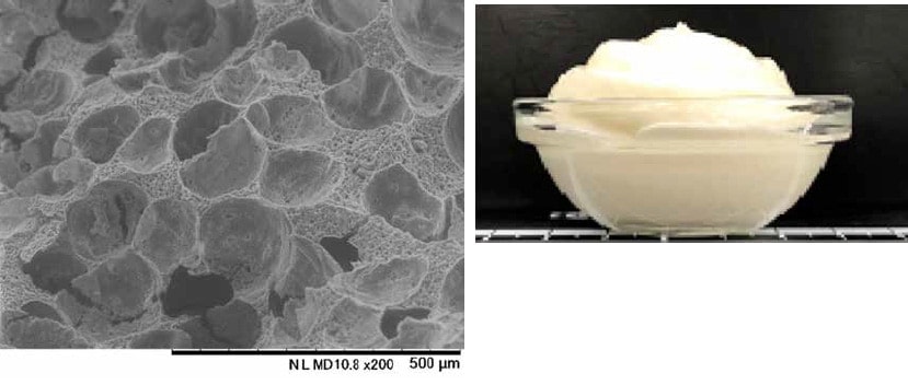
Figure 3: Espuma
Left: SEM image / Right: Sample cooking image
According to Professor Mineki, both macroscopic and microscopic observation will continue to be necessary for future developments in the field of culinary science.
“Thanks to low-vacuum SEM systems, it is now easy to observe aqueous samples. However, with black and white images, it can still be difficult to determine the composition, and artifacts from cooking and processing still get misidentified frequently. Although still rudimentary, macroscopic observations, such as taking full-view images of a sample or copies of cross sections, will continue to be essential. Researchers get a fuller understanding of the tissue images, from the macroscopic to the microscopic scale, by first identifying the morphology and material using optical and electron microscopy images. One item on my wishlist is that microscope engineers will figure out an easy way to overlay images, such as double-stained optical and SEM images.”
(Interview/article: Toshinari Yamaguchi)
References
Editor’s Notes
This interview was conducted online due to the second state of emergency declaration being issued for Covid-19. Even through the small computer screen, however, Professor Mineki’s tone conveyed her inexhaustible interest in unraveling the structure of food, and her passion for opening up the possibilities of food histology. I learned not only that the knowledge of the structure of food has helped both in advancing culinary science and developing a variety of products, but it has also improved the quality of life for the elderly and disabled. I found another ray of hope in the efforts of the researchers. I wish to thank Professor Mineki for providing a real feeling that, depending on where research is focused, the utility of low-vacuum SEM still has room to expand further.
