Kosaku Toyosaki*1
The market for high performance liquid chromatography (HPLC) has been affected by the addition of new methods for mass analysis to the range of testing methods for pharmaceuticals; this has created demand among HPLC users—particularly in the pharmaceutical industry—for mass-analysis-based structure identification. However, of the more than 200,000 LC systems in use across the worldwide HPLC market, fewer than 20% have connections to mass analyzers, creating high barriers for HPLC users to break into the world of mass analysis; in particular, at present the prevalence of mass analyzers is significantly less than that of photodetectors for HPLC applications.
Among the factors responsible for this low rate of adoption, major barriers include the installation environment required for mass analyzers, issues with maintenance and operation, and the cost of the instruments. In this report, we discuss the Chromaster 5610, a new mass detector designed specifically for the purpose of HPLC that significantly lowers the various barriers preventing HPLC users from making use of mass analysis.
Figure 1 shows a photograph of the device and lists its size and weight. The area required for the installation of this device is essentially equivalent to that required for the Chromaster series of high performance liquid chromatographs; the height of the instrument is that of 3 pump modules.
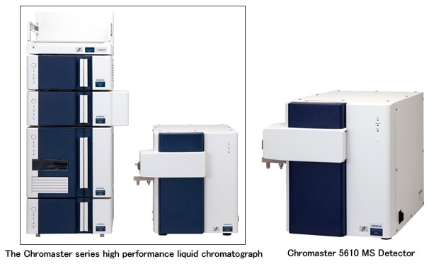
Fig.1 Hitachi Chromaster 5610 MS Detector for HPLC
Unit dimensions: 440 (W) × 610 (D) × 430 (H) mm
Unit mass: Approximately 51 kg
The key strength of this product is that it is a mass detector offering the same ease-of-use as the light-based (UV/FL) detectors typically used in HPLC, but optimized for installation in smaller spaces. In particular, the instrument offers the following advantages.
Ions produced by the ESI ion source are induced into the atmospheric ion filter (AIF) by a potential difference with the counter gas plate. The flow of ions within the atmospheric ion filter is perpendicular to the flow of ions generated in the ionization unit, helping to ensure the robustness of the apparatus. In the atmospheric ion filter, ions are separated by mobility—which is determined by charge and molecular structure—and metastable ions and cluster ions, which contribute to noise, are removed. Then, ions proceed from the AP1 into the interior of the vacuum; by displacing the axis of ion flow from the ion optical axis (the electrode axis following AP2), we prevent the accumulation of dirt on the electrode surface. Next, the ions pass through AP2, the second-stage vacuum separator, and enter Q0. Energy is uniformized through collisions with neutral molecules in Q0. Next, after passing through the three-stage vacuum separator AP3, ions enter Q1 and the subsequent analytical units. In Q1, ions to be measured are selected by the RF/DC method, in which a DC sweep is superposed on an RF signal. Ions then pass through the endcap—which performs collection for the detector—and enter the detector. The detector consists of a conversion dynode and a Channeltron® detector. In the conversion dynode, ions are exchanged for electrons; these electrons are amplified by the Channeltron® detector and detected in the form of a voltage pulse.
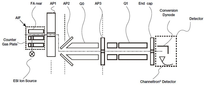
Fig.2 The ion optics system.
The ion source is a unit that ionizes sample materials flowed in from the HPLC or syringe pump (Fig. 3). The ion source in this product is compatible with electrospray ionization (ESI); in the ESI method, by applying a voltage of a few kV to counter-facing electrodes and a capillary, static liquid drops are formed, and the sample is ionized by volume compression of these liquid drops.
In this product, the sample liquid is split at a stage prior to the ionization unit and the flow rate is reduced to a few µL/min for ionization. For this reason, the quantity of nitrogen gas required during ionization may be significantly reduced. In addition, the AIF (Fig. 4) installed inside the ion source operates in ambient conditions; by applying an AC voltage with an asymmetric waveform and a variable DC voltage between two electrodes, this device separates ions based on mobility. The AIF is integrated into a single monolithic structure with the counter gas plate; maintenance may be performed simply by removing it from the ion source, with no need to stop the vacuum.
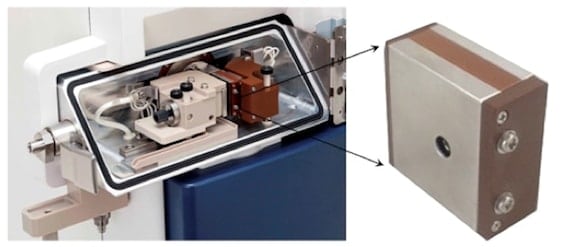
Left: Fig.3 The interior of the ion source
Right: Fig.4 The atmospheric ion filter (AIF).
Using the Chromaster 5610 MS Detector and a syringe pump, sample liquids may be inserted directly into the mass detector to obtain mass information. This allows acquisition of mass information on synthetic compounds, as well as easy monitoring of compounds while investigating synthesis conditions. Figure 5 shows a sample measurement of vildagliptin, pioglitazone, and linagliptin, compounds used as oral diabetes medications.
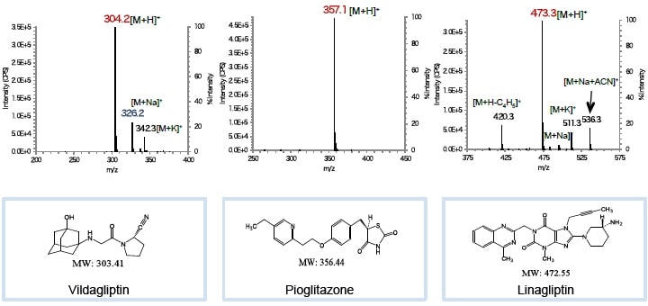
Fig.5 A sample measurement using the Chromaster 5610 MS Detector.
Chromaster 5610 MS Detector
Figure 6 shows a sample measurement involving caffeine and other methyl xanthine species. These substances are often found in coffee, cocoa products, and nutrient beverages. Because methyl xanthine species share a common basic molecular skeleton, their UV spectral patterns are very similar, and it is difficult to distinguish them based on spectra alone. However, by combining measurements with a mass detector which obtains information on substance-specific masses, the accuracy of the identification procedure is improved.
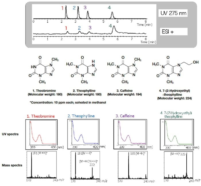
Fig.6 A sample measurement involving the LC-PDA detector and the mass detector.
Mass detector
Figure 7 shows a sample measurement involving the antibacterial agent Triclosan.
Triclosan is an antibacterial agent used against common bacteria; its applications range from health-care products such as pharmaceutical soap and toothpaste to plastics for children's toys and cloth products. Because the Chromaster 5610 MS Detector does not require any complicated configuration of settings, one obtains mass information via procedures similar to those used for UV detectors, thus obtaining calibration curves.
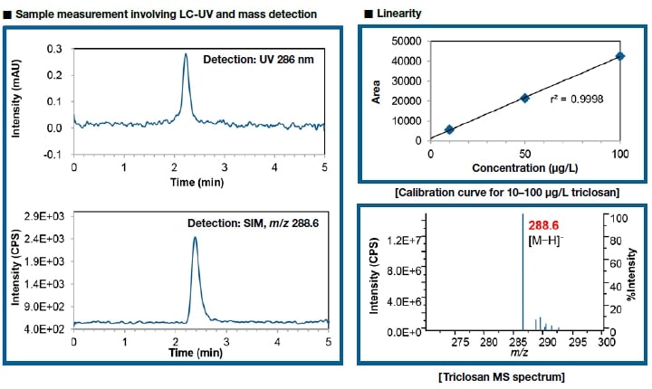
Fig.7 Sample measurement involving LC-UV and mass detection (10 µg/L triclosan)
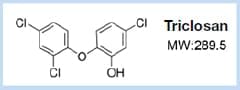
[Structure of triclosan]
Mass detector (MSD)
The Chromaster 5610 MS Detector is a new detector that avoids the problems of conventional mass analyzers that have traditionally served as barriers between HPLC users and mass-analysis techniques. The new ability to obtain mass information offers the promise of greatly enhancing the reliability of qualitative analyses.
Author
*1 Kosaku Toyosaki
Optical Instruments Design Group 1
Optical Instruments Design Department
Hitachi High-Tech Science Corporation
See more
