Akiko Wakui*1, Yasuyuki Nodera*2
When performing transmission electron microscopy (TEM) observations of samples, searching for target samples in the field of view is a particularly time-consuming task. Operators often miss a target sample when viewing samples with which they are unfamiliar or lack experience observing. This is especially true when searching for virus particles in the field of view. The Hitachi Bio-TEM series was designed to overcome these challenges. The company developed a system for automatic detection for this series of TEM instruments. The system is used on negatively stained samples that contain large amounts of impurities and items similar to the target virus, which it automatically detects.1) The Hitachi HT7800 120 kV TEM system, which is the latest model in the series, uses a suited-to-purpose digital camera to facilitate the series of tasks from observation to image acquisition.
The automatic particle search function of the HT7800 uses matching techniques and other new technologies to identify target viruses more precisely.2) This paper discusses the features and some applications of this new automatic particle search function.
The HT7800 features a maximum accelerating voltage of 120 kV and a proprietary compound objective lens. Offering both wide-field, high-contrast observations in high-contrast mode and high-resolution observation in high-resolution mode,3) the microscope is used in fields spanning from biomedicine to nanomaterials (Figure 1). The HT7800’s high-sensitivity screen camera for fluorescence observation enables observation of images of unstained samples barely perceptible to the naked eye and samples susceptible to electron beam damage, such as ice-embedded samples. The accelerating voltage can be adjusted from 20 to 120 kV to suit the sample being observed. The HT7800 also features an image navigation function that automatically images predefined locations as well as auto-focus and drift-correction functions that simplify operation.4)
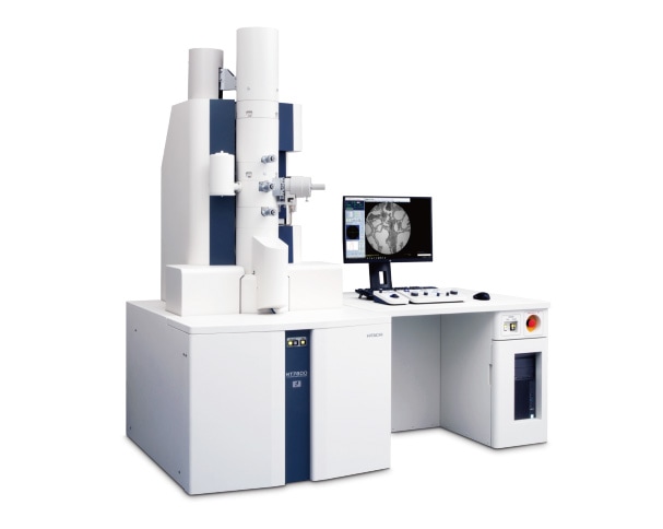
Fig. 1 Exterior of the Hitachi HT7800 120 kV TEM system.
The operating procedure for the automatic particle search function is illustrated in Figure 2. Once started, the function automatically moves the sample stage and assesses whether any target particles are present in the field of view. The function automatically records an image if particles are present or proceeds to the next field of view if no particles are present. An identification number is assigned to each particle identified in images with particles present.
Template matching is used in the automatic particle search function of the HT7800. Adjusting individual parameters allows the operator to increase particle hit rates and allows its use with certain samples that are not negatively stained.
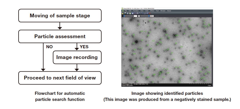
Fig. 2 Procedure for using automatic particle search function.
This study used norovirus extracted from a human stool sample. A sample suspension was applied dropwise to a 400-mesh grid with a carbon-reinforced collodion film and then negatively stained with 2% uranyl acetate. The automatic particle search function performed at an accelerating voltage of 100 kV and magnification of 20,000 × produced 36 images. The results are shown in Figure 3. Since an identification number is assigned to each norovirus particle detected in the automatically captured images (Figure 3(a)), quantitative analysis of individual particles is possible. The particle diameters and areas measured in the sample using Image Pro Premier 2D image analysis software (Media Cybernetics Inc.) are shown in Figure 3(b). The mean particle size for all detected norovirus particles and a distribution of the particle diameters can also be determined. Quantitative analysis of 4096 norovirus particles detected in this study revealed a mean particle size of 25.2 nm and a mean area of 532.3 nm2. A size distribution of the norovirus particles is shown in Figure 3(c). The distribution shows that 48% of the norovirus particles had diameters from 24 to 26 nm.
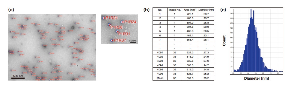
Fig. 3 Analysis of negatively stained norovirus by automatic particle search function.
(a) Particle image analysis, (b) quantitative analysis, (c) particle size distribution
The automatic particle search function of the HT7800 uses template matching. Individual template matching parameters were adjusted in an attempt to enable automatic detection of a given type of virus particle in complex tissue.
Rat cerebral cortex stained with a coronavirus (porcine hemagglutinating encephalomyelitis virus) was used as a sample. The sample was fixed with aldehyde fixing fluid and osmium tetroxide, serially dehydrated in ethanol, and embedded in an epoxy resin. The resulting sample blocks were then cut into 80-nm ultra-thin sections with an ultramicrotome. The ultra-thin sections were subjected to electronic staining with lead citrate and uranyl acetate and then observed by TEM at an accelerating voltage of 100 kV. Transmission electron micrographs of this porcine coronavirus cerebral cortex-stained sample are shown in Figure 4. One reveals an electron-dense vesicular structure in cytoplasm rich with cellular organelles such as a nucleus (N) and mitochondria (M) (Figure 4(a)). A magnified view of this image shows viral cross sections measuring 50 to 90 nm in diameter (Figure 4(b)). The automatic particle search function was used on these virus particles.
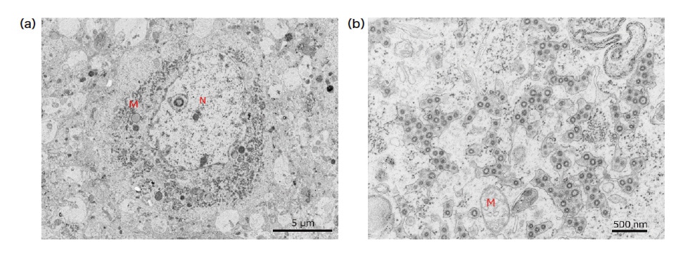
Fig. 4 Transmission electron micrographs of resin-embedded ultra-thin section of rat cerebral cortex stained with porcine coronavirus.
(a) 2500× magnification, (b) 15,000× magnification (accelerating voltage: 100 kV, N: nucleus, M: mitochondrion)
The parameters for the automatic particle search function are listed in Table 1. The parameters are set to default values experimentally determined using standard samples. For analysis of the field of view shown in Figure 5, the parameters were optimized for 90-nm coronavirus particles. This field of view is known to contain 94 virus particles. The parameters were adjusted and optimized to maximize the hit rate* of the automatic particle search function.
*Hit rate: (Number of hits / (total number of virus particles (94)) × 100
Table 1 Description of parameters
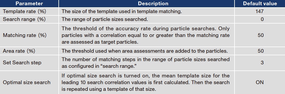
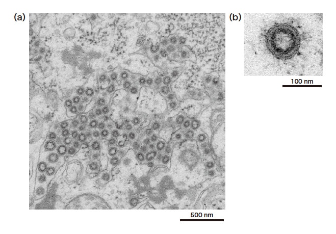
Fig. 5 Transmission electron micrograph of porcine coronavirus-stained rat cerebral cortex used to verify parameters.
(accelerating voltage: 100 kV, 10,000× magnification)
The results of verification of the search range and search steps are shown in Tables 2 and 3. The search range is a parameter for setting the particle size range. For a search range of 30% and a particle size of 90 nm, particles measuring from 63 to 117 nm are detected. As shown in Table 2, the hit rate for the default of 0% was 39%, while the hit rate increased to 66% for search ranges of 30% and 40%. A search range of 50% resulted in the highest number of detections at 81, but many misidentifications meant the hit rate was 49%. This is because a search range of 50% results in template matching from 45 to 135 nm.
Table 2 Verification of search range
Search steps represent the number of steps used for matching the size of the target particle. A greater number of steps means a higher number of matches. Hit rates were determined for 3, 5, 7, and 9 search steps. Verification was performed with a search range of 30% and automatic particle searching. As shown in Table 3, the default of 3 search steps resulted in a hit rate of 66%, while 9 search steps produced the highest hit rate of 74%. The virus particles in the sample used were cross sections and therefore had a range of sizes. This verification showed that in such samples with large particle size variance, using a larger number of search steps produces more matches.
Table 3 Verification of number of search steps
A micrograph produced by applying these optimized parameters is shown in Figure 6. Figure 6(a) shows automatically identified coronavirus particles that have been identified with a number. This demonstrates that the search function can automatically identify the target coronavirus against a complex background. The particle size distribution determined in a particle analysis of the detected coronavirus particles shows the variance in their sizes (Figure 6(b)). This large variance is attributable to the particles in the tissue section appearing as a variety of cross sections due to microtoming. Operators working with samples containing a range of particle sizes like this one are urged to optimize the search parameters so that the search function can detect the target particles with high precision and thereby improve the efficiency of TEM analysis.
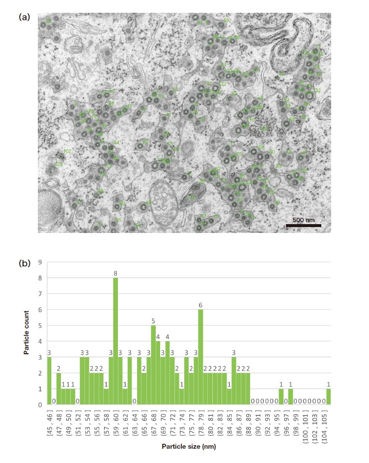
Fig. 6 Automatically detected particles in resin-embedded ultra-thin section of rat cerebral cortex stained with porcine coronavirus.
(a) Image produced by automatic particle search function (accelerating voltage: 100 kV, 15,000×magnification), (b) particle size distribution
This paper described the features and some applications of the automatic particle search function of the HT7800 TEM system. As has been discussed, the search function precisely detects the target particles in negatively stained samples and resin-embedded ultra-thin sections of tissue. TEM operators will benefit from better work efficiency and be able to precisely identify target particles with few if any misses.
References
Acknowledgments
We sincerely thank Dr. Etsuko Utagawa, Visiting Researcher, National Institute of Infectious Diseases and President, Science Lab. Yokohama, for providing the norovirus samples we used.
We also deeply thank Dr. Kinji Ishida, Institute for Biomedical Sciences, Iwate Medical University and Dr. Norio Hirano, formerly of the Department of Veterinary Microbiology, Faculty of Agriculture, Iwate University, for providing samples of rat cerebral cortex stained with coronavirus (porcine hemagglutinating encephalomyelitis virus). We are grateful for their kind contributions.
About the authors
*1 Akiko Wakui
Solution Development Dept.
Beam Technology & Analytical Systems Product Div.
Core Technology & Solutions Business Group
Hitachi High-Tech Corporation
*2 Yasuyuki Nodera
Software Design Dept.
Beam Technology & Analytical Systems Product Div.
Core Technology & Solutions Business Group
Hitachi High-Tech Corporation
