
Takashi Sekiguchi
Dr. of Science
Professor
Faculty of Pure and Applied Sciences
University of Tsukuba
Like many institutions, the University of Tsukuba has shuttered its campus and switched to online classes for all students to mitigate the spread of the novel coronavirus. In response to this situation, university faculty members are doing everything in their power to maintain the quality of their teaching—and avoid losing students who have difficulty following online courses.
However, no matter how well we may succeed in preserving the quality of our lectures, it seems essentially impossible to conduct laboratory experiments online. In particular, there is surely no substitute for the experience of turning the dials of instruments by hand, making mistakes, and devising clever workarounds for problems that arise—all essential components of an experimentalist’s training. As part of a unit on SEM techniques in a course titled Introduction to Experimental Physics and Metrology, we have recently introduced an educational innovation that—while not the same as actual experiments—offers at least a partial replica of the experimental experience, and has been well-received by our students: We use our TM4000 Plus low-vacuum tabletop microscope to observe various types of salt samples, then present students with the results of our measurements and ask them to write reports discussing the experiments. Figure 1 shows a portion of the reference material prepared for this purpose.
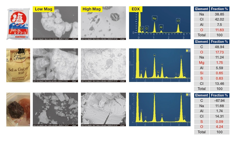
Fig. 1 SEM (backscattered-electron) images and results of EDX analysis (accelerating voltage 10 kV) for various types of salt.
Students were shown data characterizing the morphologies (backscattered-electron images) and compositions (EDX spectra and tables) of 4 types of salt—ordinary table sale, salt from the Guérande salt marshes, and pink and black Himalayan rock salt (ground using a grater)—and asked to relate these data to the tastes of the salt varieties. Significant variations in particle shapes and sizes exist even within the various salt samples, which are of varying degrees of purity and contain a smorgasbord of impurity species; a discussion of the environment in which salt crystals form can alone yield a report of several pages. In addition, the bizarre responses we received to some questions—identifying background Al as the principal component of table salt, overestimating the presence of C in EDX spectra, and allegedly detecting large quantities of organic matter contained in Guérande salt—are a source of (unintentional) hilarity. The inability to discuss results with students has forced us to grapple head-on with the difficult question of how to teach the
art of interpreting raw experimental data.
An advantage of today’s tabletop microscopes is the availability of low-vacuum operating modes, which allow samples to be observed without conductive coatings. For SEM in particular, this has been nothing short of a revolutionary advance, instantly broadening the range of observable samples. Although tabletop microscopes at the University of Tsukuba are shared-use facilities, with the exception of ferromagnetic nanoparticles there are no restrictions on the solid samples that may be observed. However, this fact seems not to be widely appreciated; in practice, most researchers in their 40s or older continue to believe that SEM observation of insulators requires the application of coatings—and, moreover, that moisture-containing materials and other observation samples that might damage instruments are to be avoided at all costs. In contrast, younger generations of researchers—who learned the technique after the advent of low-vacuum SEM—feel unencumbered by limitations on SEM observations; at worst one encounters absent-minded students who might carelessly introduce liquids into the sample chamber.
The advantage of low-vacuum tabletop microscopy is that ideas for new experiments can be implemented as soon as they are conceived: with no need to apply coatings, sample preprocessing consumes little time. Here we exploit this feature to address a question of particularly timely importance: whether face masks are effective in preventing the spread of the novel coronavirus. This topic has recently been debated in various forums, including morning TV programs; however, no matter how persuasive an argument one might hear for the ineffectiveness of masks made from non-woven fabrics, truly convincing proof remains elusive. Thus, immediately upon arriving at our laboratory we conducted an experiment in which an airflow containing micron-sized nylon particles was passed through facemasks and the results observed in our TM4000 system. As shown in Figure 2, disposable masks made of non-woven cloth have a three-layer structure, consisting of a layer of fine-pored non-woven cloth sandwiched between two layers of coarse-pored non-woven cloth. Eureka! Our experiments show that the fine-pored cloth layer traps 1 μm particles, while larger particles stick to the adhesion region of the outer coarse-pored layer, perhaps due to static electricity. Unfortunately, particles of sizes below 1 μm were not readily available, and thus our work has not yet produced any answers regarding virus behavior; nonetheless, we have learned a considerable amount about the role of masks. For example, one conclusion that will surely remain valid, come what may, is this: when masks are reused, one must take care to distinguish between front and back!
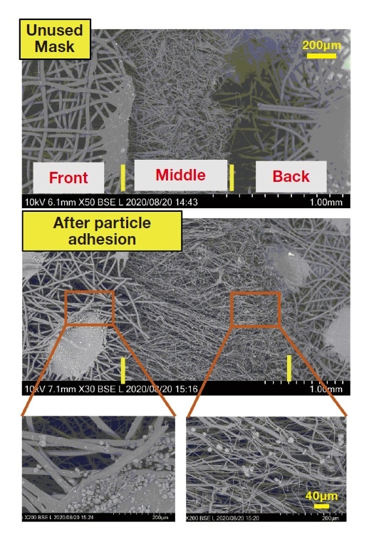
Fig. 2 SEM images of a non-woven cloth mask. Upper panel: New, unused mask. Center panel: After particle adhesion. Lower panels: Enlargements of center-panel image. These are backscattered-electron images acquired at an accelerating voltage of 10 kV.
Our final examples involve salt crystals and spiders. Two years ago, several massive typhoons made landfall in Japan, causing major destruction even in regions such as Chiba and Ibaragi that rarely experience natural disasters. In Tsukuba, the morning after one major storm passed through the region I awoke to a sunny day with clear post-typhoon skies—but automobile hoods felt somehow sticky when touched, bringing my attention to the fact that the typhoon dropped a host of other improbable things by gale-force winds. It was right around the time that we found a spider web on the side of our campus building, and decided to sample a portion of it for observation in the TM4000 Plus, with the results shown in Figure 3. According to our dictionary of zoology, the vertical strands of spiderwebs consist of twisted bundles of strings, while the horizontal strands consist of single strings dotted with drops of a sticky substance. It seems our sampling procedure did not distinguish between the two types of strands, and thus our experimental sample consisted of a tangle of both. SEM (backscattered-electron) images (Figure 3, left) revealed white spots on the strands, which we analyzed in greater detail via EDX element mapping, yielding the color images shown at the right of Figure 3. The white spots show up as bright regions in Na and Cl maps, indicating that they are salt crystals. Such fanciful notions—not only that a typhoon might carry seawater all the way to Tsukuba, which lies 100 km from the ocean, but also that salt particles in the water might crystallize and adhere to the strands of spiderwebs—seemed too farfetched for us even to have imagined! Prior to the formation of these crystals, salt condensed in rain particles, yielding salty raindrops that wound up sticking to spiderwebs. Needless to say, our thoughts turned immediately to the poor spiders, whom we hoped would avoid contracting high blood pressure from drinking so much salty water.
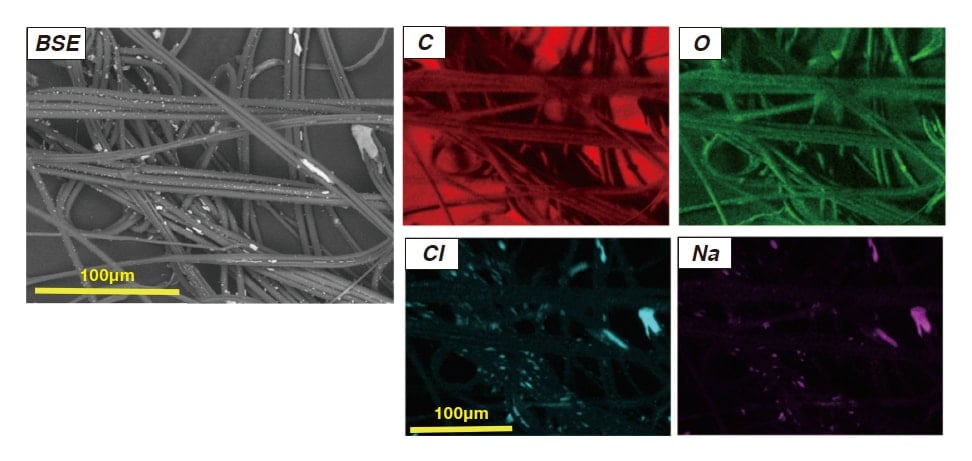
Fig. 3 Strands of a spiderweb sampled the day after a typhoon. Left: SEM (backscattered-electron) image, captured at accelerating voltage of 10 kV. Right: X-ray elemental mapping images.
And now back to our subject. Low-vacuum SEM systems are simple instruments without high-vacuum exhaust systems that have expanded the SEM user base; these systems eliminate the need for sample coating and other timeconsuming pre-processing steps, and we expect they will eventually become as ubiquitous in laboratories as optical microscopes are today. To add just one final thought, we have noticed that there is some empty space behind the door of the sample chamber of our TM system; perhaps, with some ingenuity, it might be possible to use this space for current-supplying wires, or sample heaters, or manipulatable stages, or other innovations to create new measurement environments? In any event, I am hopeful that simple, user-friendly low-vacuum SEM systems will become more and more common in the years ahead, bringing the power of SEM observation to a broader community of users.
Notes
See more
