


Microscopy & Microanalysis 2026 (M&M 2026)
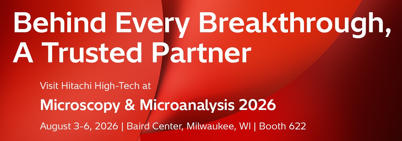
Join us at Microscopy & Microanalysis 2026 (M&M 2026) and discover the latest innovations and technologies Hitachi offers. Whether you are currently manufacturing or developing next generation systems, you will learn about state-of-the-art equipment and analytical techniques.
Available for demonstration or discussion will be solutions of automation, mineralogy, battery development/inspection, and advanced sample preparation.
Lunch Talks and Vendor Tutorials at Booth 622*
At M&M 2026, Hitachi High-Tech will offer a series of tutorials and presentations including Lunch Talk and after-hour Vendor Tutorial sessions. Do not miss this opportunity to learn about the latest technologies, collaborations, and innovations by customers.
* Food & drink will be served. Limited capacity. First come, first served.
** Schedule and topics may change.

Monday, August 3rd
 Reserve your spot |
Lunch Talk (12:00 PM – 12:45 PM) Environmental transmission electron microscopy from UHV to near atmospheric pressure Dr. Frances M. Ross, Massachusetts Institute of Technology Department of Materials Science and Engineering DetailsEnvironmental transmission electron microscopy (ETEM) offers prospects for understanding fundamental phenomena and answering real-world questions by visualizing materials transformations with high spatial and temporal resolution. I will present two strategies we find useful to optimize the experimental design and broaden the range of problems we can address: ultra-high vacuum techniques to quantify reactions in environmentally sensitive materials, and sample geometries that access higher pressures while still enabling the surface-sensitive secondary electron signal to be collected. With these and other continuing experimental developments, ETEM is well placed to expand its impact in addressing key materials problems with uniqueness and precision.BioFrances M. Ross is a faculty member at the Department of Materials Science and Engineering at the Massachusetts Institute of Technology in Cambridge, MA, USA. She received her B.A. in Physics and Ph.D. in Materials Science from Cambridge University, UK, and along the way became an enthusiast of electron microscopy. She extended her interests to include in situ microscopy during her postdoc at A.T.&T. Bell Laboratories, then as a Staff Scientist at the National Center for Electron Microscopy, Lawrence Berkeley National Laboratory, and finally as a Research Staff Member at the IBM T. J. Watson Research Center, before joining MIT. Her research is based around the development of in situ electron microscopy techniques to help understand crystal growth, epitaxy, self-assembly and electrochemical and other liquid phase processes. |
 Reserve your spot |
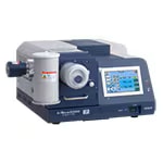
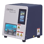
Vendor Tutorial (5:45 PM – 6:45 PM) From Sample to Insight: End-to-End Automated Mineralogy with Hitachi AMICS and GeoMineral AI Insights Jonathan P. Knapp, Ph.D, Director of New Business Development DetailsAutomated mineralogy has transformed how we quantify mineralogy, liberation, and particle characteristics. However, bottlenecks persist at both ends of the workflow: preparing granular samples and turning analytical output into actionable insight. This tutorial will present an integrated approach from Hitachi High-Tech that addresses the full chain. We begin with a new automated sample preparation system for granular and particulate samples, standardizing mounting and polishing to reduce operator variability and substantially increase throughput. We then introduce the next generation of AMICS software, featuring redesigned particle analysis workflows aligned with ISO standards for reproducible, audit-ready results. Finally, we unveil GeoMineral AI Insights, a data delivery portal that surfaces AMICS results through AI-assisted querying, visualization, and reporting, placing interpretation in the hands of decision-makers rather than instrument operators alone. Join us to see how Hitachi is closing the loop from sample to insight. |
Tuesday, August 4th
 Reserve your spot |
Lunch Talk (12:00 PM – 12:45 PM) Live High-Throughput Imaging Using Automated Workflows with Hitachi’s Latest HT7800II TEM Heather Berensmann, Application Engineer DetailsIntroducing Hitachi’s newly released 120 kV transmission electron microscope, the HT7800II TEM combines high-quality, reproducible data acquisition with improved efficiency to support a diverse range of applications. With enhanced automation capabilities, the HT7800II now supports automated workflow recipes using Hitachi’s EM Flow Creator software. Join us for a real-time demonstration on how the HT7800II with EM Flow Creator can streamline TEM workflows and support high-throughput, high precision data collection. |
 Reserve your spot |
Tutorial (5:45 PM – 6:45 PM Booth 1504) Strategies for Applying 4D-STEM and Ptychography from 20 to 300 keV Dr. Arthur M. Blackburn, Department of Physics and Astronomy and Centre for Advanced Materials and Related Technologies, University of Victoria, Canada DetailsFour-Dimensional Scanning Transmission Electron Microscopy (4D-STEM) and electron ptychography are transforming electron microscopy by allowing atomic resolution insights from low energy (≤ 30 keV) scanning electron microscopy (SEM) which rivals or complements the information obtainable from much higher energy aberration corrected instruments. For example, a resolution of at least 0.67 Å has recently been demonstrated using ptychography with a 20 keV electron beam operating in a transmission mode using a SEM with a cold emission gun and immersion lens1. This was realized through combination of adding a diffraction projector lens to our SEM; using an un-coated hybrid direct electron detector2; and incorporating correction of diffraction projector distortions3 within a multi-slice ptychographic reconstruction process. Here I will discuss the strategies we have used to practically collect and process 4D-STEM data for a range of applications and analyses, including ptychography, within our research facility. Applying these techniques to data collected in smaller and lower energy SEM type instruments reduces the need for observations traditionally performed in higher energy instruments and thus widens the access to atomic resolution insights.1Blackburn, A. M., Cordoba, C., et al, Nature Communications 16, 8977 (2025); 2Tinti, G., Marchetto, H. et al., Journal of Synchrotron Radiation 24, 963-974 (2017); 3Fitzpatrick, M. R. C., Blackburn, A. M., et al., Ultramicroscopy 283, 114347 (2026) BioArthur Blackburn holds the Hitachi High-Tech Canada Research Chair in Advanced Electron Microscopy at the University of Victoria, where he directs the Advanced Microscopy Facility and is an Associate Professor in the Department of Physics and Astronomy. Prior to joining the University of Victoria, he was a Senior Research Scientist in the Hitachi Cambridge Laboratory, embedded with the Cavendish Laboratory of the University of Cambridge. He progressed to this role after completing his PhD at the University of Cambridge, Department of Physics. |
Wednesday, August 5th
 Reserve your spot |
Lunch Talk (12:00 PM – 12:45 PM) Fix, Conduct, and Quantify: Next-Gen Tissue Fixation for Analytical and Quantitative Volume Electron Microscopy Eduardo Rosa-Molinar, Ph.D., Professor, Cell Biology and Physiology; Professor, Neuroscience; Scientific Director, WashU Center for Cellular Imaging; Faculty, Division of Biology and Biomedical Sciences; Member, Brain Tumor Center at Siteman Cancer Center; Member, Institute of Clinical and Translational Sciences; Faculty, Imaging Science, McKelvey School of Engineering, WashU in Saint Louis School of Medicine DetailsAre sample charging and structural degradation limiting your Analytical and Quantitative volume Electron Microscopy (AQvEM) data? Join us to discover a breakthrough in sample preparation. This talk introduces a patented conductive fixative designed specifically for high-fidelity analytical and quantitative AQvEM.Traditional methods often compromise ultrastructural preservation to achieve conductivity. This newly patented formulation extends tissue protection while eliminating charging artifacts. Learn how this technology enables superior, artifact-free 3-D reconstruction and precise quantification in challenging biological systems, including brain tissue, brain tumors, and organoids. See how you can transform your tissue workflows from qualitative imaging to precise analytical quantification. BioEduardo Rosa-Molinar, Ph.D., is a principal investigator track, tenured Professor of Cell Biology and Physiology (primary appointment), principal investigator track, tenured Professor of Neuroscience, Faculty of Imaging Science, and the Scientific Director of Washington University Center for Cellular Imaging at the Washington University in Saint Louis School of Medicine. Until July 1, 2024, he served as Director of Microscopy and Analytical Imaging and as a tenured Professor of Pharmacology, Toxicology, Neuroscience, and Bioengineering at the University of Kansas.He holds the lifetime distinction of Fellow of the American Association for the Advancement of Science (AAAS). He serves as Trustee of the Biological Stain Commission. He also holds memberships and fellowships in numerous societies and serve or served in various senior leadership positions, committees, and boards, including the Society for Integrative Biology, Histochemical Society, Royal Microscopical Society, Linnean Society of London, Optica (formally Optical Society of America), Institute of Electrical an Electronics Engineers (IEEE), Society for Neuroscience, American Society of Cell Biology, Institute of Physics, Society of Photographic Instrumentation Engineers, Association of Biomolecular Facilities, Biological Stain Commission, BioImaging North America, Volume Electron Microscopy Community (vEM), Correlated Multi-modal Imaging in Life Sciences (COMULIS), Quality Assessment and Reproducibility for Instruments and Images in Light Microscopy (QUAREP-LiMi), Microscopy Society of America, Australian Microscopy and Microanalysis Society, Microanalysis Society, International Microphysiological Systems Society, International Society For Stem Cell Research, Metastasis Research Society, American Association for Cancer Research, Society for Neuro-Oncology, International Cancer Imaging Society, Society of Nuclear Medicine and Molecular Imaging (SNMMI), Missouri Academy of Science, and the Federation of American Societies of Experimental Biology. He has also served as review editor, associate editor, and editorial board member of various journals, including Frontiers in Molecular Neuroscience, IEEE Access, Current Protocols in Imaging, Biotechnic and Histochemistry, Integrative and Comparative Biology, and Journal of Experimental Zoology Part B: Molecular and Developmental Evolution. |
Instruments Available for Demos
The following instruments will be available for private demos. Please reserve your spot in advance.
NEW! TEM: HT7800II 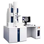 
|
SEM: SU3900 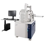
|
FE-SEM: SU8700 UHR 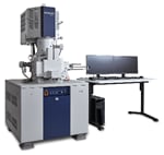
|
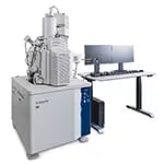
|
Automated Material Characterization: AMICS 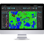
|
|
|
|
First time at M&M: New! HT7800II 120 kV TEM
[Speed and Reliability, Delivered]
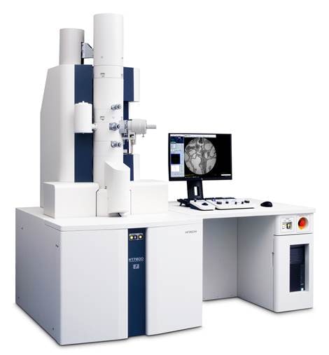
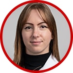
Hitachi’s new HT7800II 120 kV TEM combines high-quality, reproducible data acquisition with improved efficiency to support a diverse range of applications across multiple fields. With added workflow automation as standard, streamline TEM routine alignments automatically with a single click. Improve screening capabilities to search with precision by capturing Whole View images of the entire grid within 10 seconds. Accelerate productivity by methodically collecting images and montages at specified locations throughout the grid to reshape TEM data acquisition. Discover more by capturing what matters with the new HT7800II TEM.
- Special Promotion available! Learn more here.
- Don’t miss the Lunch Talk on the New HT7800II on Tuesday, August 4th! Reserve your seat in advance.
- Private demos are available. Reserve your demo time.
Dates
August 3-6, 2026
Location
Booth 622, Baird Center, Milwaukee, WI
Contact
Hitachi High-Tech America, Inc.
Event info and registration