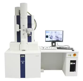
Transmission Electron Microscope HT7800 Series
Speed and Reliability, Delivered
The HT7800 series is a 120 kV transmission electron microscope (TEM) that combines high-quality and reproducible data acquisition with improved efficiency for observation work.
By automating routine alignments and settings, and featuring an intuitive design that streamlines operations from field-of-view search to image capture, it supports high quality data acquisition regardless of users expertise.
The HT7800 series contributes to enhancing productivity in research and inspection tasks across diverse fields, from medical and life science to materials development.
Features
-
ACHIEVE MORE, WITH EASE:
Access reliable data effortlessly, no matter the experience level.
SMART WORKFLOW:
Enhanced automation capabilities to streamline and reduce labor during the TEM data acquisition process
UNLIMITED SCOPE:
Diverse applications spanning a wide range of fields in science and technology
-
Hitachi's proprietary dual-mode objective lens provides versatility by supporting a wide variety of applications. Perform two different lens actions to achieve both High Contrast (HC) and High Resolution (HR) imaging capabilities with a single click. Low magnification provides easy navigation across the entire grid, High Contrast (HC) observation allows increased contrast over a large field of view for life science capabilities, and High Resolution (HR) observation reduces the focal length for enhanced resolution for materials science capabilities. The HT7800 series is the only system in the industry to offer both high-contrast and high-resolution expandability without compromise.
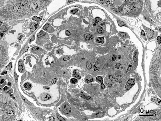
High-contrast observation
Specimen: Rat kidney
Accelerating voltage: 80 kV; Magnification: x300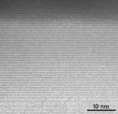
High-resolution observation
Specimen: Asbestos (Crocidolite)
Accelerating voltage: 120 kV; Magnification: x400,000 -
The HT7800 series offers an ergonomic workstation for a fully-integrated workflow in one intuitive guided user interface. Both image navigation, using the wide field-of-view screen camera, and image acquisition, using the high-resolution camera, are carried out in one software platform. Digitalized operations also benefit remote applications, some of which include simultaneously sharing and exchanging captured TEM images at multiple points.
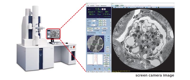
High-sensitivity screen camera allows intuitive operation
-
Combining an intuitive workflow with sophisticated automation capabilities allows users of any experience level to achieve high-throughput, high-quality data acquisition. The system features enhanced automation capabilities, including automatic beam alignments to support routine alignment tasks, Image Navigation for automated and efficient image acquisition, and EM Flow Creator*1 for flexible workflow configuration.
*1Option
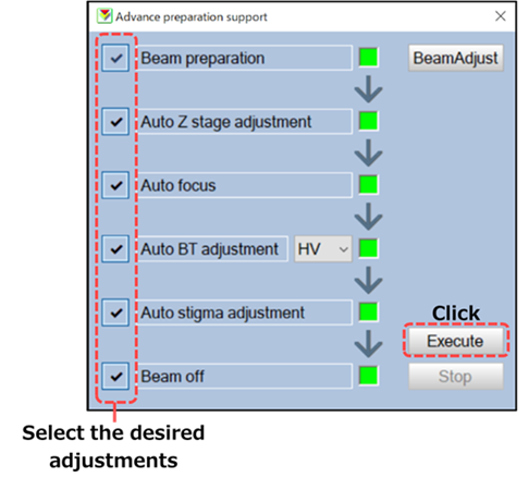
Automatic Beam Alignment*2
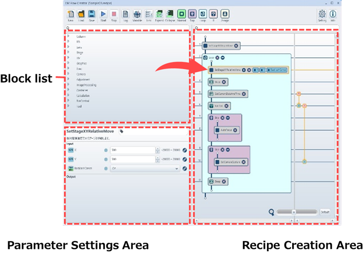
EM Flow Creator
*2Auto Z stage adjustment requires the optional 5-axis stage.
-
The HT7800 series comes standard with various features such as image-navigation capabilities to automate image acquisition, as well as wide-area automatic image capture, 3D tomography, and more. The system also supports a variety of optional capabilities, including a CLEM system*1, STEM*1, EDS elemental analysis*1 and Cryo observation*1.
*1Option
Specification
| HT7800II | HT7820 | HT7830 | |
|---|---|---|---|
| Electron gun | W, LaB6 | ||
| Accelerating voltage | 20 - 120 kV (100 V/step variable) | ||
| Resolution (Lattice) | 0.20 nm (Off-axis, 100 kV) |
0.14 nm (Off-axis, 120 kV) |
0.14 nm (Off-axis, 120 kV) 0.19 nm (On-axis, 120 kV) |
| Maximum magnification | x600,000 | x800,000 | x1,000,000 |
| Stage maximum tilt angle | ±70° | ±30° | ±10° |
| Standard features | Automatic beam alignment, Auto focus, Microtrace, Autodrive, Live FFT display, Measurement function, Low dose, API (auto pre-irradiation), Image navigation function, Column with mild baking function, Whole view function, Drift correction function etc. | ||
Application Data
Life science
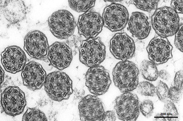
Cilia
Model : HT7800, Acceleration voltage : 120 kV, Magnification : x20,000
In the cross-section of the cilia, a microtubule pattern known as the 9+2 structure can be observed.
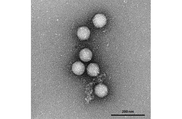
Adenovirus (Negative staining)
Model : HT7820, Acceleration voltage : 120 kV, Magnification : x40,000
The surface structures of the adenoviruses are explicitly observed.
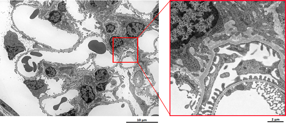
Mouse Kidney (5x5tiling)
Model : HT7800, Acceleration voltage : 80 kV, Magnification : x5,000
By using tiling, it is possible to acquire wide-field, high-resolution data.
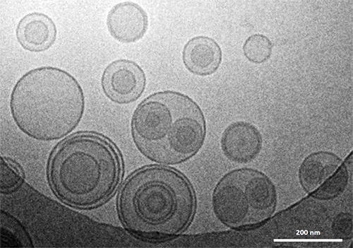
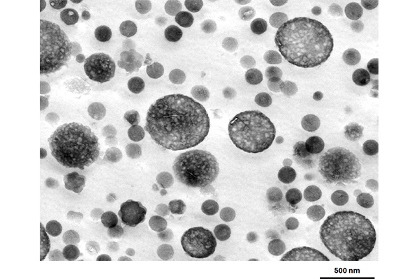
Liposome (Cryo observation)
Model : HT7800Ⅱ, Acceleration voltage : 120 kV, Magnification : x30,000
The shape and membrane structure of the liposomes are clearly visible.
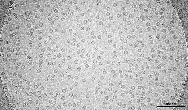
Virus-like particles (Cryo observation)
Model : HT7800, Acceleration voltage : 100 kV, Magnification : x70,000
Stable cryo observation is possible.
Material
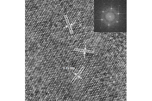
Pyrophyllite
Model : HT7800Ⅱ, Acceleration voltage : 120 kV, Magnification : x600,000
Even in pyrophyllite, which is sensitive to electron beams, a three-directional lattice pattern (0.45 nm) can be observed.
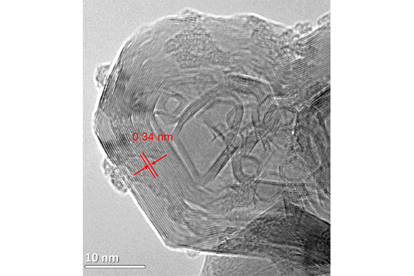
Carbon graphite
Model : HT7820, Acceleration voltage : 120 kV, Magnification : x800,000
A clear lattice fringe of graphite with a d-spacing of approximately 0.34 nm is observed.
ABS
Model : HT7800Ⅱ, Acceleration voltage : 100 kV, Magnification : x20,000
The butadiene rubber in the ABS is clearly visible as a salami-type structure.
Semiconductor
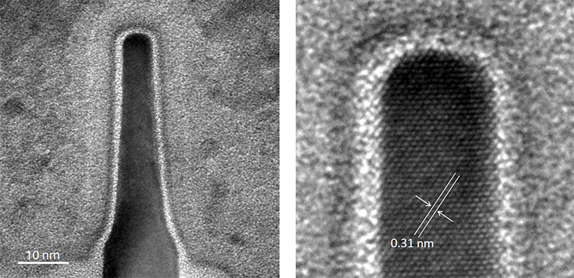
Fin-FET
Model : HT7830, Acceleration voltage : 120 kV, Magnification : x600,000
A clear lattice image of silicon with a spacing of approximately 0.31 nm is observed.
The Application Data Collection has a "Life Science" section and a "Materials and Semiconductors" section, introducing typical observation data and application examples in each field.
Citations
 Powered by Bioz
This journal addresses a wide range variety of research papers and useful application data using Hitachi science instruments.
Photo collections of beauty of metals, minerals, organisms etc. reproduced by the electron microscope and finished more beautifully by computer graphic technology.
Related Product Categories
Related Information

Hitachi High-Tech Social Media