
Tabletop Microscopes TM4000PlusIII/TM4000III
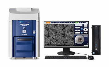
A tabletop microscope for the next generation of scientific leaders.
The TM4000PlusIII/TM4000III is the newest addition to a lineup of microscopes that have sold over 5,800 units worldwide.
Our newest updates bring improvements to users at the forefront of R&D, quality control, and education.
Features
-
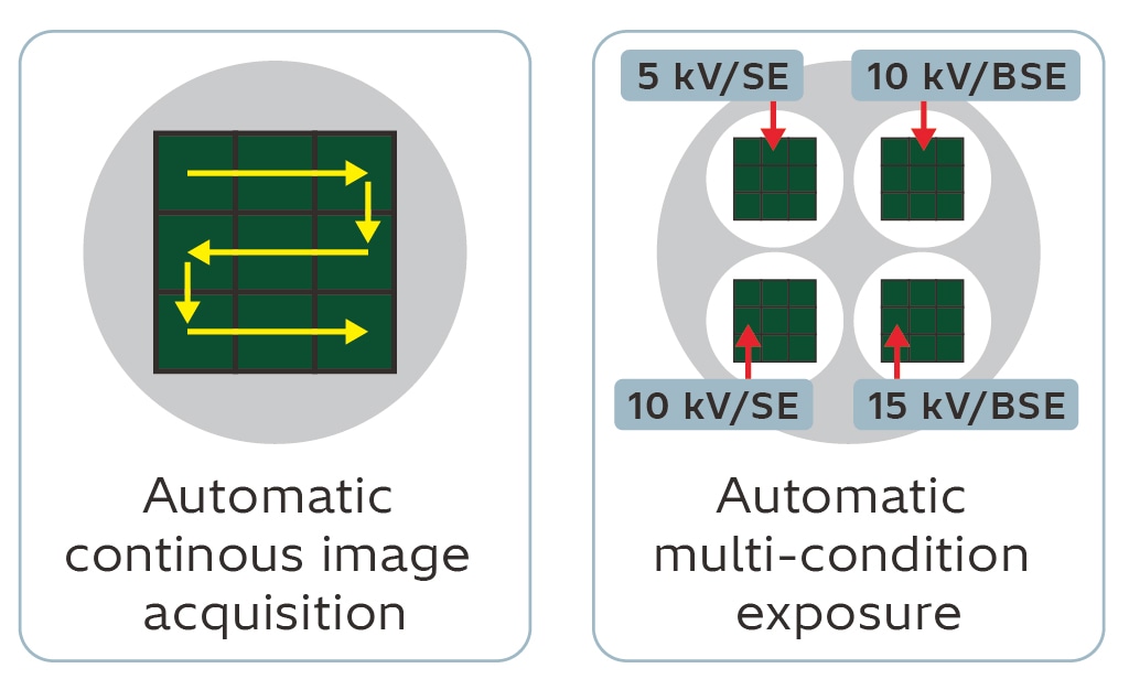
Automated observation of multiple specimens: Recipe Control*1
The TM4000PlusIII recipe function can be used to automatically execute operations, such as stage movement, magnification setting, and image capturing. Once the user builds a recipe, it can be executed to run the tool automatically with no further intervention. This significantly reduces the workload on operators and allows for more consistent results, even from inexperienced users.
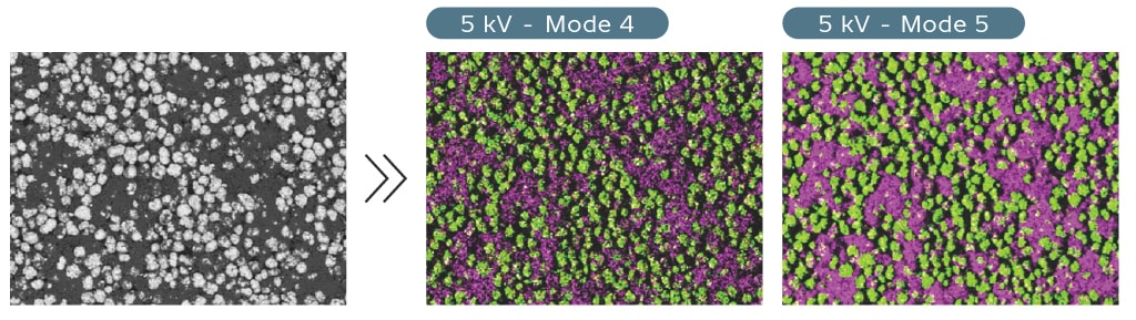
Faster automatic particle analysis: High-current function + AZtecLiveLite particle analysis package*
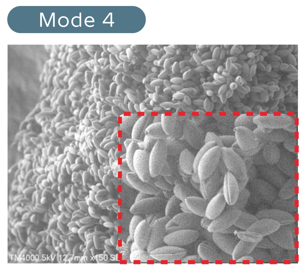
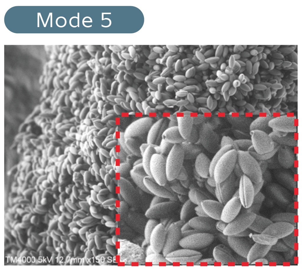
Tasks such as purity analysis of industrial products and particle analysis of filters can be time consuming as data is required from many locations to complete the analysis. The TM4000PlusIII is equipped with a new high-current setting, Mode 5, which provides increased signal for imaging and EDS analysis. In combination with the Oxford Instruments EDS AZtecLiveLite particle analysis package, this helps to increase throughput.
Field-of-view navigation with less noise: High-current function
SEM field-of-view navigation is performed using fast scanning modes, so images tend to be prone to noise. The high-current function allows for easy field-of-view navigation due to its high signal-to-noise (S/N) ratio. This takes the stress out of specimen navigation!
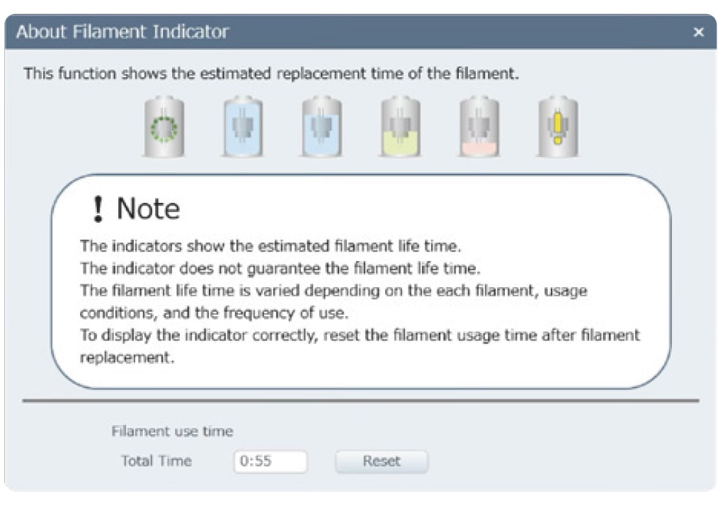
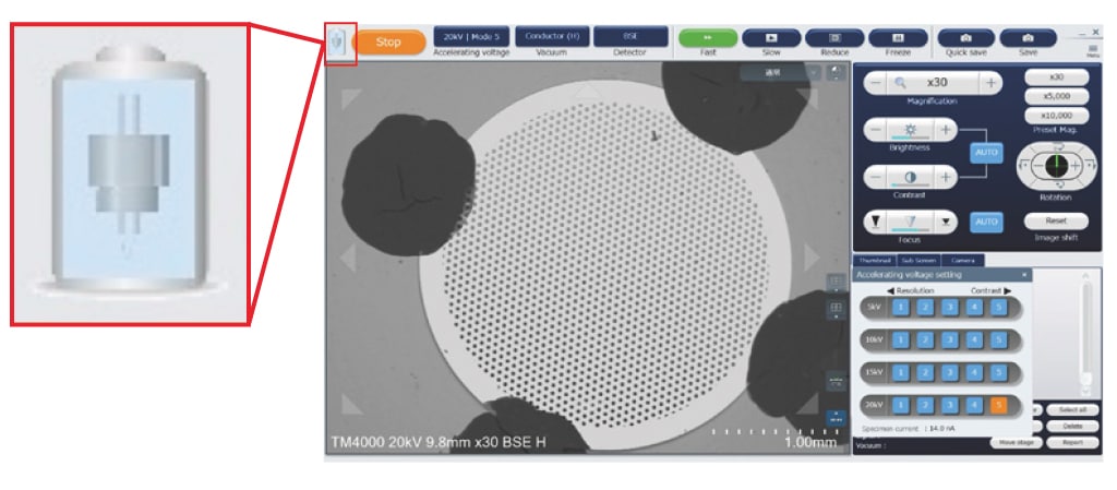
Plan Ahead with the Filament Monitoring Function
Maintenance planning: Filament monitoring function
The TM4000PlusIII is equipped with a system that monitors the filament and displays graphics to show the remaining lifetime of the filament. If the equipment is being operated by multiple users, this makes it possible to plan the use of the tool in a systematic way. There is no more need to worry about the filament suddenly running out when the tool is most needed.
Ideal for Educational Purposes
Experience the world of electron microscopy with no preprocessing required:
Low-vacuum function + high-sensitivity backscattered-electron detectorUsers can try out electron microscopy with all kinds of specimens: picked flowers, treasured minerals, or familiar foods, with no need for metal coating. The low-vacuum function of the TM4000PlusIII/TM4000III reduces preprocessing, and the high-sensitivity backscattered-electron detector (BSED) allows images to be obtained quickly.
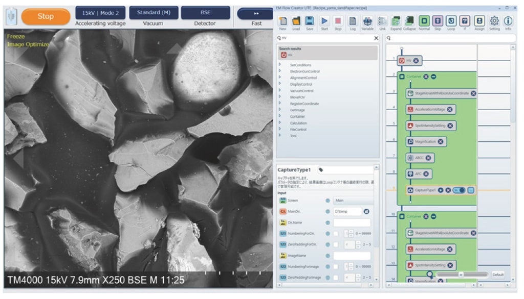
Build programming skills with workflow automation functions (TM4000PlusIII)*1
Developing and maintaining digital skills is vital in today’s world. The automation support function allows you to experientially learn important programming concepts such as "sequential execution," "repetition," and "conditional branching" through the operation of the TM4000PlusIII.
*1 Optional for TM4000PlusIII only
*2 Optional
-
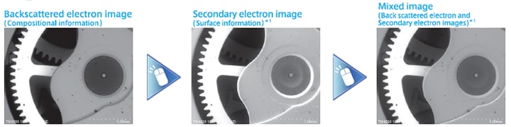
Automation, Observation, and Elemental Analysis
Easily obtain images from different detectors with a single click: SE, BSE, or Mixed Signal.
Rapid acquisition of elemental maps *2
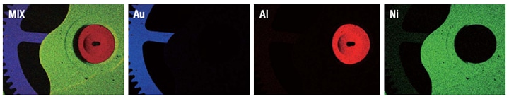
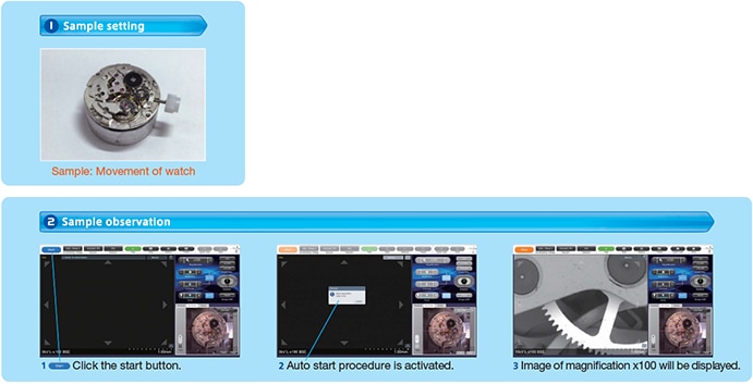
Sample: Wrist watch
*1 Secondary electron images and MIX images can only be observed in TM4000Plus III
*2 Option
Intuitive operation on Camera Navi
Camera Navi provide optical correrlation to SEM stage positions for easy navigation to areas of interest.
SEM images can be overlayed on the SEM MAP image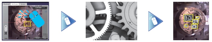
Sample: Wrist watch
Report Creator*
Easily select images and templates to create customized reports.
Reports can be saved/edited in Microsoft Office® formats.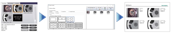
Sample: Wrist watch
* Option
-
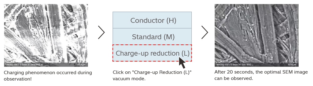
Charge-up reduction mode
Vacuum adjustments, to reduce sample charging, can be done with a single click.
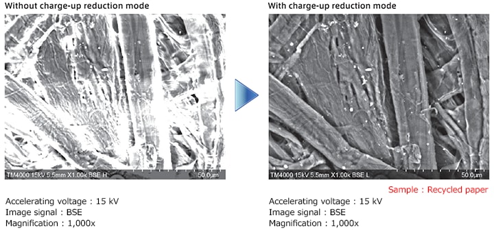
Image a variety of materials under low-vacuum condition
Utilize a variety of low-vacuum conditions to image different materials.
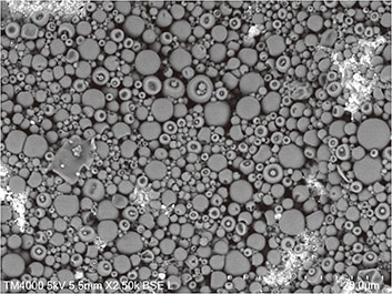
Sample: Paint ink
Accelerating voltage: 5 kV
Image signal: BSE
Magnification: 2,500x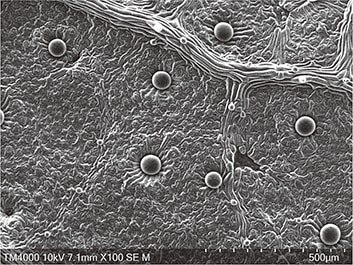
Sample: Leaf of plant
Accelerating voltage: 10 kV
Image signal: SE
Magnification: 100x -
Innovative secondary-election detector obtains surface detail with non-conductive samples at lower vacuum conditions
The TM4000PlusIII can observe not only conductive samples, but also non-conductive or hydrated samples without extensive sample preparation. Users can switch between BSE and SE signals easily.
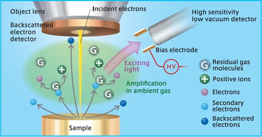
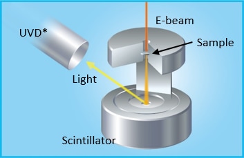
High-sensitivity Low-vacuum SE Detector (UVD)
Hitachi's UVD generates secondary-electron images from visible light through electron-gas interactions.
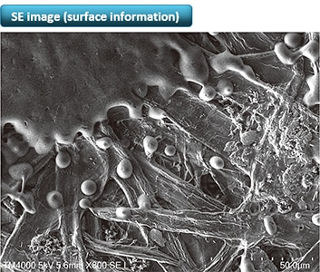
Accelerating voltage: 5 kV
Image signal: SE
Magnification: 800x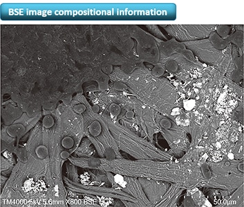
Sample: Printed paper
Accelerating voltage: 5 kV
Image signal: BSE
Magnification: 800x -
High-accelerating voltage enables higher-speed EDS analysis.
EDS mapping data at 20 kV in 2 min
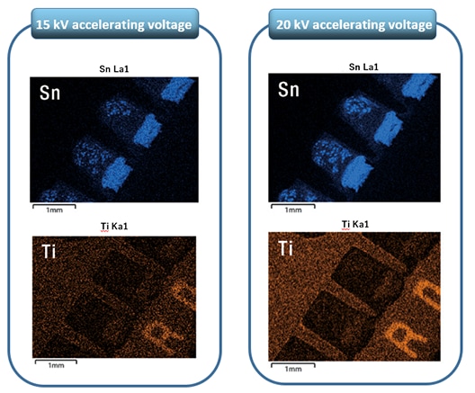
Sample: Electronic components -
Multi Zigzag automatically capture multiple SEM images and stitch together into a single high-resolution image.
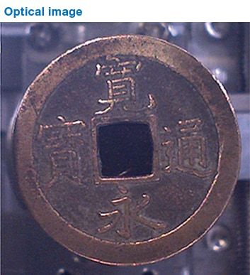
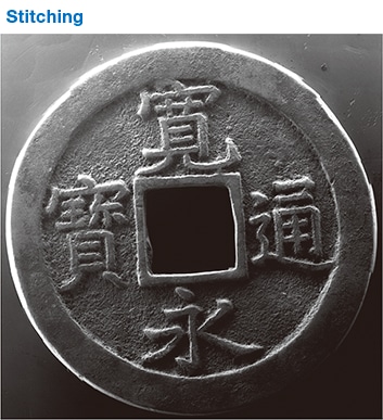
Sample: Japanese ancient coin
Accelerating voltage: 15 kV
Image signal: SE
Magnification: 30x
Field of view 10 vertically × 12
horizontally
(some parts were trimmed)* Option
-
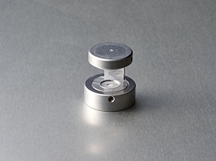
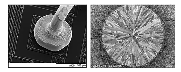
STEM holder (Option)
Easily obtain transmitted images on thin samples
The newly developed STEM holder is used to perform transmission images in combiantion with the Hitachi UVD.
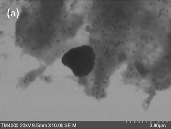
Sample: Abrasive
Accelerating voltage: 20 kV
Image signal: (a) STEM, (b) BSE
Magnification: 10,000 x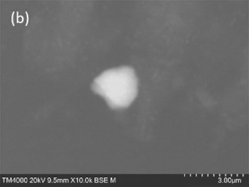
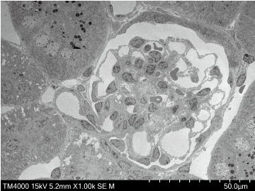
Sample: Rat kidney
Accelerating voltage: 15 kV
Image signal: STEM
Magnification: 1,000 x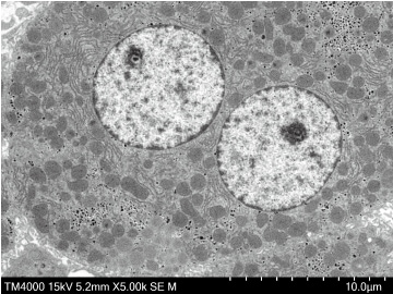
Sample: Rat liver
Accelerating voltage: 15 kV
Image signal: STEM
Magnification: 5,000 x* Option
* UVD is an optional function for the TM4000III
Applications Gallery
Specifications
| Model name | TM4000Plus III | TM4000 III |
|---|---|---|
| Magnifications |
10× - 100,000× (Photographic magnification) 25× - 250,000× (monitor display magnification) |
|
| Accelerating voltage | 5 kV, 10 kV, 15 kV, 20 kV | |
| Image signal | Backscattered electron Secondary electron Mix (Backscattered electron + Secondary electron) |
Backscattered electron |
| Vacuum mode | Conductor: BSE Standard: BSE/SE/Mixed Charge-up reduction: BSE/SE/Mixed |
Standard Charge-up reduction |
| Sample stage traverse | X: 40 mm, Y: 35 mm | |
| Maximum sample size | 80 mm (diameter), 50 mm (thickness) | |
| Electron gun | Pre-centered cartridge tungsten filament | |
| Signal detection system | High-Sensitivity 4-segment BSE detector High-Sensitivity Low-Vacuum SE detector (UVD) |
High-Sensitivity 4-segment BSE detector |
| Evacuation system (vacuum pump) |
Turbo molecular pump : 67 L/s×1 unit Diaphragm pump : 20 L/min×1 unit |
|
| Stage | Motorized stage | Manual stage |
| Size / weight | Main unit330 (width)×614 (depth)× 547 (height) mm 55 kg |
Main unit: 330 (width)×617 (depth)× 547 (height) mm 53 kg |
| Diaphragm pump: 144 (width)×270 (depth)×216 (height) mm, 5.5kg | ||
 Powered by Bioz