
Analysis Example of Aflatoxin M1
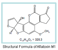
Food-contaminating mycotoxins include aflatoxin, ochratoxin, deoxynivalenol/nivalenol, and patulin. Aflatoxin is toxic to the liver and is said to be the strongest natural carcinogen. Aflatoxin B1, B2, G1, and G2 are naturally occurring, and B1 is a strong carcinogen. It is known that aflatoxin B1 is partly metabolized to aflatoxin M1 in the body. Therefore, when a dairy cow eats fodder contaminated with aflatoxin B1, aflatoxin M1 may be excreted into milk.
There is also a movement to establish an official method in Japan and the Evaluation Committee of Mycotoxin Methodology is investigating the method.
Presented here is the analysis of aflatoxin M1 by an HPLC-fluorescence detection method, which is method 2 described under "GB 5413.37-2010: Determination of Aflatoxin M1 in Milk and Milk Products," National Food Safety Standard of the People's Republic of China.
Regulation Values of Aflatoxin M1 in Milk and Milk Products in Each Countries
| Japan*1 under review | China*2 | U.S.*3 | |
|---|---|---|---|
| Regulation value | NMT 0.5 µg/kg | NMT 0.5 µg/kg | NMT 0.5 µg/kg |
*1 Collaborative Study Protocol by the Evaluation Commitee of Mycotoxin Methodology "Analysis Method for Aflatoxin M1 in Milk"
*2 Method 2, GB 5413.37-2010: Determination of Aflatoxin M1 in Milk and Milk Products
*3 AOAC Official Method 2000.8, Aflatoxin M1 in Liquid Milk, Immumoaffinity Column by Liquid Chromatography.
Analysis Example of Aflatoxin M1
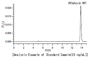
Analytical Conditions
| Column | HITACHI LaChrom C18 (5 µm) (4.6 mm I.D. x 250 mm) |
|---|---|
| Eluent | Acetonitrile/Water = 25/75 (v/v) |
| Flow rate | 1.0 mL/min |
| Column temperature | 30°C |
| Detection wavelength | FL Ex 365 nm, Em 435 nm |
| Injection vol. | 10 µL |
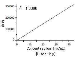
A good linearity with a coefficient of determination of 1.000 was obtained over a range of 0.1 - 40 ng/mL.
The lower detection limit was less than 0.02 ng, which is less than the lower detection limit described in the GB, which confirms sufficient sensitivity. In addition, when the measurement was repeated 6 times at 20 ng/mL, the peak retention time reproducibility (%RSD) and peak area reproducibility (%RSD) were respectively 0.064% and 0.80%, indicating good reproducibility.
NOTE:
These data are an example of measurement; the individual values cannot be guaranteed.
The system is for research use only, and is not intended for any animal or human therapeutic or diagnostic use.
In order to read a PDF file, you need to have Adobe® Reader®