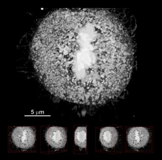
Three Dimensional Structure of the HeLa Cell
© Sumire Inaga (Tottori University, Faculty of Medicine)
Three dimensionally reconstructed images of a cultured HeLa cell stained with platinum blue are presented; the images are composed of cross-section images serially acquired with 10 nm steps using an FIB-SEM.
Morphologies of chromosomes or cytoplasmic processes and their dispersion states in the cell are clearly visualized. It is expected that higher-order structure of unknown chromosomes will be elucidated by this technique.
1st Prize. At 73th photo contest hosted by the Japanese Society of Microscopy in 2017.
Condition
Specimen : Cultured HeLa cell
Instrument: Focused Ion & Electron Beam System nanoDUE'T NB5000
Magnification : X 4,000
Accelerating voltage : 3 kV (BSE)
*This work was presented at the "photo contest" hosted by the Japanese Society of Microscopy.
*Reproduction or republication without permission prohibited.
*"nanoart" is registered trademark of Hitachi High-Tech Corporation in Japan.
Products & Services
Related Information

Hitachi High-Tech Social Media