Flowers on the lake
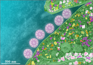
This is the pseudo color picture of dark field STEM image of a cell body of the protozoan parasites on the mouse duodenum and its line hair section by a scanning transmission electron microscope. Distribution of 9+2 structure of microtubule of line hair and ribosome cytoplasm granules seems to be six aquatic plants blooming in a flower garden and an inlet opening in a quiet shore. Such beautiful micro world structure reminds of a mystery of life.
At 61st photo contest hosted by the Japanese Society of Microscopy in 2005.
Condition
Specimen: Mouse Duodenum buried in resin
Instrument: Ultra-thin Film Evaluation System HD-2300
Magnification: × 50,000
Accelerating voltage: 200 kV
*All information related to these photographers is based on the information when the photo was taken.
*This work was presented at the "photo contest" hosted by the Japanese Society
of Microscopy.
*Reproduction or republication without permission prohibited.
*"nanoart" is registered trademark of Hitachi High-Tech Corporation in Japan.
Products & Services
Related Information

Hitachi High-Tech Social Media